当前位置:
X-MOL 学术
›
Nat. Protoc.
›
论文详情
Our official English website, www.x-mol.net, welcomes your feedback! (Note: you will need to create a separate account there.)
Nanoscale imaging of clinical specimens using conventional and rapid-expansion pathology.
Nature Protocols ( IF 14.8 ) Pub Date : 2020-04-01 , DOI: 10.1038/s41596-020-0300-1 Octavian Bucur 1, 2, 3, 4 , Feifei Fu 5 , Mike Calderon 6 , Geetha H Mylvaganam 7, 8 , Ngoc L Ly 7, 8 , Jimmy Day 9 , Simon Watkin 6 , Bruce D Walker 7, 8, 10 , Edward S Boyden 9, 11, 12, 13, 14, 15 , Yongxin Zhao 5, 9
Nature Protocols ( IF 14.8 ) Pub Date : 2020-04-01 , DOI: 10.1038/s41596-020-0300-1 Octavian Bucur 1, 2, 3, 4 , Feifei Fu 5 , Mike Calderon 6 , Geetha H Mylvaganam 7, 8 , Ngoc L Ly 7, 8 , Jimmy Day 9 , Simon Watkin 6 , Bruce D Walker 7, 8, 10 , Edward S Boyden 9, 11, 12, 13, 14, 15 , Yongxin Zhao 5, 9
Affiliation
|
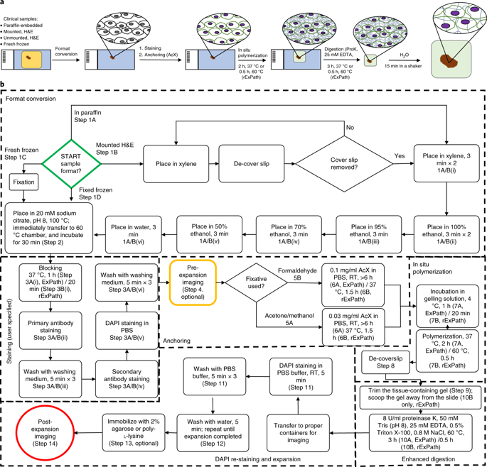
In pathology, microscopy is an important tool for the analysis of human tissues, both for the scientific study of disease states and for diagnosis. However, the microscopes commonly used in pathology are limited in resolution by diffraction. Recently, we discovered that it was possible, through a chemical process, to isotropically expand preserved cells and tissues by 4-5× in linear dimension. We call this process expansion microscopy (ExM). ExM enables nanoscale resolution imaging on conventional microscopes. Here we describe protocols for the simple and effective physical expansion of a variety of human tissues and clinical specimens, including paraffin-embedded, fresh frozen and chemically stained human tissues. These protocols require only inexpensive, commercially available reagents and hardware commonly found in a routine pathology laboratory. Our protocols are written for researchers and pathologists experienced in conventional fluorescence microscopy. The conventional protocol, expansion pathology, can be completed in ~1 d with immunostained tissue sections and 2 d with unstained specimens. We also include a new, fast variant, rapid expansion pathology, that can be performed on <5-µm-thick tissue sections, taking <4 h with immunostained tissue sections and <8 h with unstained specimens.
中文翻译:

使用常规和快速扩展病理学对临床标本进行纳米级成像。
在病理学中,显微镜是用于分析人体组织的重要工具,无论是用于疾病状态的科学研究还是用于诊断。但是,病理学中常用的显微镜的分辨率受到衍射的限制。最近,我们发现可以通过化学过程使保存的细胞和组织各向同性地扩展线性尺寸4-5倍。我们称这种过程扩展显微镜(ExM)。ExM可以在常规显微镜上进行纳米级分辨率成像。在这里,我们描述了各种人类组织和临床标本(包括石蜡包埋,新鲜冷冻和化学染色的人类组织)的简单有效物理扩展的协议。这些协议只需要廉价的,常规病理实验室中常见的市售试剂和硬件。我们的协议是为有常规荧光显微镜经验的研究人员和病理学家编写的。常规方法,扩展病理学,可以在约1天内用免疫染色的组织切片完成,而在2天内用未染色的标本完成。我们还包括一种新的,快速变化的,快速扩展的病理学,可以在厚度小于5 µm的组织切片上进行,免疫染色的组织切片需要4个小时,未染色的样本需要8个小时。
更新日期:2020-04-01
中文翻译:
使用常规和快速扩展病理学对临床标本进行纳米级成像。
在病理学中,显微镜是用于分析人体组织的重要工具,无论是用于疾病状态的科学研究还是用于诊断。但是,病理学中常用的显微镜的分辨率受到衍射的限制。最近,我们发现可以通过化学过程使保存的细胞和组织各向同性地扩展线性尺寸4-5倍。我们称这种过程扩展显微镜(ExM)。ExM可以在常规显微镜上进行纳米级分辨率成像。在这里,我们描述了各种人类组织和临床标本(包括石蜡包埋,新鲜冷冻和化学染色的人类组织)的简单有效物理扩展的协议。这些协议只需要廉价的,常规病理实验室中常见的市售试剂和硬件。我们的协议是为有常规荧光显微镜经验的研究人员和病理学家编写的。常规方法,扩展病理学,可以在约1天内用免疫染色的组织切片完成,而在2天内用未染色的标本完成。我们还包括一种新的,快速变化的,快速扩展的病理学,可以在厚度小于5 µm的组织切片上进行,免疫染色的组织切片需要4个小时,未染色的样本需要8个小时。


 京公网安备 11010802027423号