当前位置:
X-MOL 学术
›
Chem. Mater.
›
论文详情
Our official English website, www.x-mol.net, welcomes your
feedback! (Note: you will need to create a separate account there.)
Artificial Blood Vessel Frameworks from 3D Printing-Based Super-Assembly as In Vitro Models for Early Diagnosis of Intracranial Aneurysms
Chemistry of Materials ( IF 7.2 ) Pub Date : 2020-03-30 , DOI: 10.1021/acs.chemmater.0c00208 Rongke Gao 1 , Xin Tian 1 , Qizheng Li 1 , Xuefei Song 1 , Bing Shao 2 , Jie Zeng 3 , Zhanjie Liu 4 , Debo Zhi 5 , Gang Zhao 5 , Hongming Xia 4 , Bensheng Qiu 5 , Guang Chen 6 , Kang Liang 7 , Pu Chen 6 , Dongyuan Zhao 3 , Biao Kong 3
Chemistry of Materials ( IF 7.2 ) Pub Date : 2020-03-30 , DOI: 10.1021/acs.chemmater.0c00208 Rongke Gao 1 , Xin Tian 1 , Qizheng Li 1 , Xuefei Song 1 , Bing Shao 2 , Jie Zeng 3 , Zhanjie Liu 4 , Debo Zhi 5 , Gang Zhao 5 , Hongming Xia 4 , Bensheng Qiu 5 , Guang Chen 6 , Kang Liang 7 , Pu Chen 6 , Dongyuan Zhao 3 , Biao Kong 3
Affiliation
|
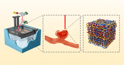
Intracranial aneurysm (IA) is a bulge from the weak area in the wall of cerebral blood vessels and can cause serious diseases, such as hemorrhagic stroke and other neurologic diseases. Experimental and computational results demonstrated that the different flow fields of blood had a great influence on the formation, growth, and rupture of IAs. Therefore, it is crucial to acquire flow field of blood for fully characterizing the hemodynamics. In this study, six transparent models of artificial blood vessels with different growth stages of IAs by 3D printing-based super-assembly technology were first fabricated. Epoxy-based resin was used to form a 3D pipeline structure, and it played an important role in restoring the appearance of IAs and comparing with the medical image. Phase contrast-magnetic resonance imaging (PC-MRI) and computational fluid dynamics (CFD) were used to assess flow fields of IA during growth. The internal flow and wall shear stress (WSS) of inner IAs showed a very low level in the cardiac cycle compared with normal blood vessels. The CFD and PC-MRI demonstrated that the internal flow of IA gradually interfered with intravascular flow because IAs formed, and this interference gradually reduced after a mid-term stage. Meanwhile, the growth and rupture points of side IAs mainly located in the efferent region of IAs may result from the blood flow becoming extremely slow in this area. This proposed 3D printing-based super-assembly technology reduced the replica size by at least 80% and provided a visual internal structure to obtain MR imaging data.
中文翻译:

基于3D打印的超级组件的人工血管框架作为颅内动脉瘤的早期诊断的体外模型
颅内动脉瘤(IA)是脑血管壁薄弱部位的隆起,可引起严重的疾病,例如出血性中风和其他神经系统疾病。实验和计算结果表明,不同的血液流场对IAs的形成,生长和破裂有很大的影响。因此,获取血液的流场以充分表征血液动力学至关重要。在这项研究中,通过基于3D打印的超级装配技术,首先制造了六个透明的具有不同IAs生长阶段的人造血管模型。环氧树脂基树脂用于形成3D管道结构,并且在恢复IA的外观以及与医学图像进行比较中起着重要作用。相衬磁共振成像(PC-MRI)和计算流体动力学(CFD)用于评估IA在生长过程中的流场。与正常血管相比,内部IA的内部流量和壁切应力(WSS)在心动周期中显示出非常低的水平。CFD和PC-MRI显示,由于IAs的形成,IA的内部流量逐渐干扰了血管内流量,并且在中期之后这种干扰逐渐减少。同时,主要位于IAs传出区域的侧面IAs的生长和破裂点可能是由于该区域的血流变得极其缓慢所致。这项基于3D打印的超级组装技术的提出将复制品的尺寸减少了至少80%,并提供了可视内部结构来获取MR成像数据。
更新日期:2020-04-23
中文翻译:
基于3D打印的超级组件的人工血管框架作为颅内动脉瘤的早期诊断的体外模型
颅内动脉瘤(IA)是脑血管壁薄弱部位的隆起,可引起严重的疾病,例如出血性中风和其他神经系统疾病。实验和计算结果表明,不同的血液流场对IAs的形成,生长和破裂有很大的影响。因此,获取血液的流场以充分表征血液动力学至关重要。在这项研究中,通过基于3D打印的超级装配技术,首先制造了六个透明的具有不同IAs生长阶段的人造血管模型。环氧树脂基树脂用于形成3D管道结构,并且在恢复IA的外观以及与医学图像进行比较中起着重要作用。相衬磁共振成像(PC-MRI)和计算流体动力学(CFD)用于评估IA在生长过程中的流场。与正常血管相比,内部IA的内部流量和壁切应力(WSS)在心动周期中显示出非常低的水平。CFD和PC-MRI显示,由于IAs的形成,IA的内部流量逐渐干扰了血管内流量,并且在中期之后这种干扰逐渐减少。同时,主要位于IAs传出区域的侧面IAs的生长和破裂点可能是由于该区域的血流变得极其缓慢所致。这项基于3D打印的超级组装技术的提出将复制品的尺寸减少了至少80%,并提供了可视内部结构来获取MR成像数据。


 京公网安备 11010802027423号