Kidney International ( IF 14.8 ) Pub Date : 2020-03-28 , DOI: 10.1016/j.kint.2020.02.024 Satoshi Saeki 1 , Hiroyuki Ohba 2 , Yuko Ube 1 , Kayoko Tanaka 1 , Waka Haruyama 1 , Masako Uchii 1 , Tetsuya Kitayama 1 , Hideo Tsukada 2 , Takashi Shimada 1

|
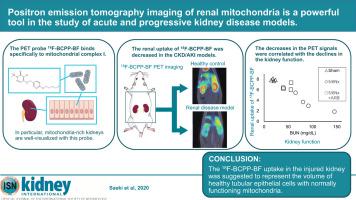
Mitochondrial dysfunction plays a critical role in the pathogenesis of kidney diseases via ATP depletion and reactive oxygen species overproduction. Nonetheless, few studies have reported the renal mitochondrial status clinical settings, partly due to a paucity of methodologies. Recently, a positron emission tomography probe, 18F-BCPP-BF, was developed to non-invasively visualize and quantitate the renal mitochondrial status in vivo. Here, 18F-BCPP-BF positron emission tomography was applied to three mechanistic kidney disease models in rats: kidney ischemia-reperfusion, 5/6 nephrectomy and anti-glomerular basement membrane glomerulonephritis. In rats with ischemia-reperfusion, a slight decrease in the kidney uptake of 18F-BCPP-BF was accompanied by morphological abnormality of the mitochondria in the proximal tubular cells after three hours of reperfusion, when the kidney function was slightly declined. In 5/6 nephrectomy and rats with anti-glomerular basement membrane glomerulonephritis, the kidney uptake of 18F-BCPP-BF cumulatively decreased with impairment of the kidney function, which was accompanied by a reduction of mitochondrial protein and a pathological tubulointerstitial exacerbation rather than glomerular injury. The 18F-BCPP-BF uptake in the injured kidney was suggested to represent the volume of healthy tubular epithelial cells with normally functioning mitochondria. Thus, this positron emission tomography probe can be a powerful tool for studying the pathophysiological meanings of the mitochondrial status in kidney disease.
中文翻译:

肾脏线粒体的正电子发射断层扫描成像是研究急性和进行性肾脏疾病模型的有力工具。
线粒体功能障碍通过ATP耗竭和活性氧过量产生,在肾脏疾病的发病机理中起着关键作用。尽管如此,很少有研究报道肾脏线粒体状态的临床状况,部分原因是缺乏方法学。最近,开发了一种正电子发射断层扫描探头18 F-BCPP-BF,用于在体内无创地观察和定量肾脏线粒体的状态。在这里,将18 F-BCPP-BF正电子发射断层扫描应用于大鼠的三种机制性肾脏疾病模型:肾脏缺血-再灌注,5/6肾切除术和抗肾小球基底膜肾小球肾炎。在缺血再灌注大鼠中,肾脏摄取18F-BCPP-BF再灌注3小时后,肾功能略有下降,并伴有近端肾小管细胞线粒体形态异常。在5/6肾切除术和患有抗肾小球基底膜肾小球肾炎的大鼠中,肾脏对18 F-BCPP-BF的摄入累积减少,但肾功能受损,这伴随着线粒体蛋白的减少和病理性肾小管间质加重而不是肾小球损伤。在18建议在受伤的肾脏中摄取F-BCPP-BF代表线粒体功能正常的健康肾小管上皮细胞的体积。因此,这种正电子发射断层扫描探头可以成为研究肾脏疾病中线粒体状态的病理生理意义的有力工具。


 京公网安备 11010802027423号