Scientific Reports ( IF 3.8 ) Pub Date : 2020-03-27 , DOI: 10.1038/s41598-020-62633-4 Tzu-Yu Hou , Tung-Mei Kuang , Yu-Chieh Ko , Yu-Fan Chang , Catherine Jui-Ling Liu , Mei-Ju Chen

|
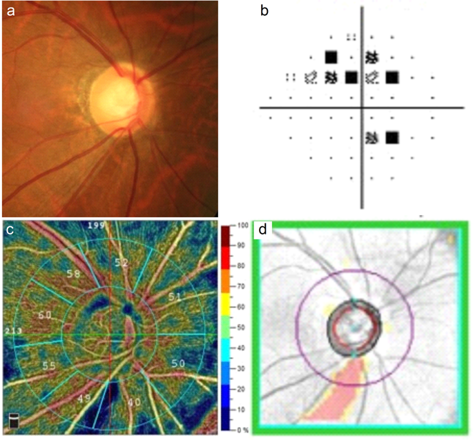
There is distinct pathogenesis between primary open-angle glaucoma (POAG) and primary angle-closure glaucoma (PACG). Although elevated intraocular pressure (IOP) is the major risk factor for glaucoma, non-IOP risk factors such as vascular abnormalities and lower systolic/diastolic perfusion pressure may play a role in the pathogenic process. This study aimed to compare the vessel density (VD) in the optic disc and macula using optical coherence tomography angiography (OCTA) between POAG and PACG eyes. Thirty-two POAG eyes, 30 PACG eyes, and 39 control eyes were included. All the optic disc VD parameters except the inside disc VD were significantly lower in glaucomatous eyes than in control eyes. Compared with PACG eyes, only the inferior temporal peripapillary VD was significantly lower in POAG eyes. The parafoveal VD was significantly lower in each quadrant in glaucomatous eyes than in control eyes. The central macular and parafoveal VD did not differ between POAG and PACG eyes. In conclusion, the inferior temporal peripapillary VD was significantly reduced in POAG eyes compared with PACG eyes, while PACG eyes showed a more evenly distributed reduction in the peripapillary VD. The distinct patterns of VD change may be associated with the different pathogenesis between POAG and PACG.
中文翻译:

通过光学相干断层扫描血管造影测量开角型和闭角型青光眼的视盘和黄斑血管密度。
原发性开角型青光眼(POAG)和原发性闭角型青光眼(PACG)之间有不同的发病机制。尽管眼内压(IOP)升高是青光眼的主要危险因素,但血管异常和收缩压/舒张压灌注压降低等非眼内压危险因素可能在发病过程中发挥作用。本研究旨在使用光学相干断层扫描血管造影 (OCTA) 比较 POAG 和 PACG 眼的视盘和黄斑中的血管密度 (VD)。包括 32 只 POAG 眼、30 只 PACG 眼和 39 只对照眼。除内视盘 VD 外,青光眼眼的所有视盘 VD 参数均显着低于对照眼。与PACG眼相比,POAG眼仅下颞部视乳头周围VD显着降低。青光眼眼每个象限的中心凹 VD 均显着低于对照眼。POAG 和 PACG 眼的中央黄斑和中心凹 VD 没有差异。总之,与PACG眼相比,POAG眼的下颞部视乳头周围VD显着减少,而PACG眼的视乳头周围VD减少分布更均匀。VD 变化的不同模式可能与 POAG 和 PACG 不同的发病机制有关。


 京公网安备 11010802027423号