当前位置:
X-MOL 学术
›
Pediatr. Res.
›
论文详情
Our official English website, www.x-mol.net, welcomes your
feedback! (Note: you will need to create a separate account there.)
State-of-the-art neonatal cerebral ultrasound: technique and reporting
Pediatric Research ( IF 3.1 ) Pub Date : 2020-03-01 , DOI: 10.1038/s41390-020-0776-y Jeroen Dudink 1 , Sylke Jeanne Steggerda 2 , Sandra Horsch 3, 4 ,
Pediatric Research ( IF 3.1 ) Pub Date : 2020-03-01 , DOI: 10.1038/s41390-020-0776-y Jeroen Dudink 1 , Sylke Jeanne Steggerda 2 , Sandra Horsch 3, 4 ,
Affiliation
|
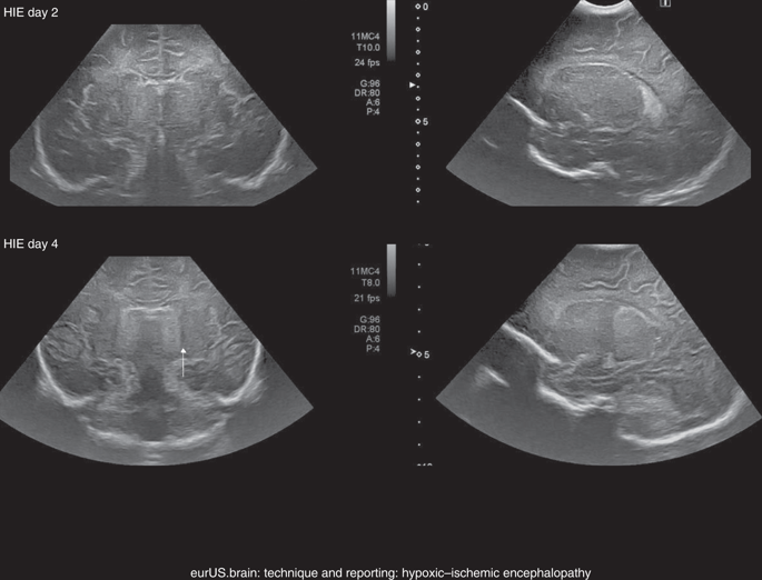
In the past three decades, cerebral ultrasound (CUS) has become a trusted technique to study the neonatal brain. It is a relatively cheap, non-invasive, bedside neuroimaging method available in nearly every hospital. Traditionally, CUS was used to detect major abnormalities, such as intraventricular hemorrhage (IVH), periventricular hemorrhagic infarction, post-hemorrhagic ventricular dilatation, and (cystic) periventricular leukomalacia (cPVL). The use of different acoustic windows, such as the mastoid and posterior fontanel, and ongoing technological developments, allows for recognizing other lesion patterns (e.g., cerebellar hemorrhage, perforator stroke, developmental venous anomaly). The CUS technique is still being improved with the use of higher transducer frequencies (7.5–18 MHz), 3D applications, advances in vascular imaging (e.g. ultrafast plane wave imaging), and improved B-mode image processing. Nevertheless, the helpfulness of CUS still highly depends on observer skills, knowledge, and experience. In this special article, we discuss how to perform a dedicated state-of-the-art neonatal CUS, and we provide suggestions for structured reporting and quality assessment.
中文翻译:

最先进的新生儿脑超声:技术和报告
在过去的三年中,脑超声 (CUS) 已成为研究新生儿大脑的可靠技术。这是一种相对便宜的、非侵入性的、床边神经影像学方法,几乎在每家医院都可用。传统上,CUS 用于检测主要异常,例如脑室内出血 (IVH)、脑室周围出血性梗塞、出血后脑室扩张和(囊性)脑室周围白质软化 (cPVL)。使用不同的声窗,例如乳突和后囟门,以及持续的技术发展,可以识别其他病变模式(例如,小脑出血、穿支中风、发育性静脉异常)。CUS 技术仍在随着更高换能器频率 (7.5–18 MHz) 的使用、3D 应用、血管成像的进步(例如 超快平面波成像),以及改进的 B 模式图像处理。尽管如此,CUS 的帮助仍然高度依赖于观察者的技能、知识和经验。在这篇特别文章中,我们讨论了如何进行专门的最先进的新生儿 CUS,并为结构化报告和质量评估提供建议。
更新日期:2020-03-01
中文翻译:
最先进的新生儿脑超声:技术和报告
在过去的三年中,脑超声 (CUS) 已成为研究新生儿大脑的可靠技术。这是一种相对便宜的、非侵入性的、床边神经影像学方法,几乎在每家医院都可用。传统上,CUS 用于检测主要异常,例如脑室内出血 (IVH)、脑室周围出血性梗塞、出血后脑室扩张和(囊性)脑室周围白质软化 (cPVL)。使用不同的声窗,例如乳突和后囟门,以及持续的技术发展,可以识别其他病变模式(例如,小脑出血、穿支中风、发育性静脉异常)。CUS 技术仍在随着更高换能器频率 (7.5–18 MHz) 的使用、3D 应用、血管成像的进步(例如 超快平面波成像),以及改进的 B 模式图像处理。尽管如此,CUS 的帮助仍然高度依赖于观察者的技能、知识和经验。在这篇特别文章中,我们讨论了如何进行专门的最先进的新生儿 CUS,并为结构化报告和质量评估提供建议。


 京公网安备 11010802027423号