当前位置:
X-MOL 学术
›
Pediatr. Res.
›
论文详情
Our official English website, www.x-mol.net, welcomes your
feedback! (Note: you will need to create a separate account there.)
Preterm white matter injury: ultrasound diagnosis and classification
Pediatric Research ( IF 3.1 ) Pub Date : 2020-03-01 , DOI: 10.1038/s41390-020-0781-1 Thais Agut 1 , Ana Alarcon 1 , Fernando Cabañas 2 , Marco Bartocci 3 , Miriam Martinez-Biarge 4 , Sandra Horsch 5, 6 ,
Pediatric Research ( IF 3.1 ) Pub Date : 2020-03-01 , DOI: 10.1038/s41390-020-0781-1 Thais Agut 1 , Ana Alarcon 1 , Fernando Cabañas 2 , Marco Bartocci 3 , Miriam Martinez-Biarge 4 , Sandra Horsch 5, 6 ,
Affiliation
|
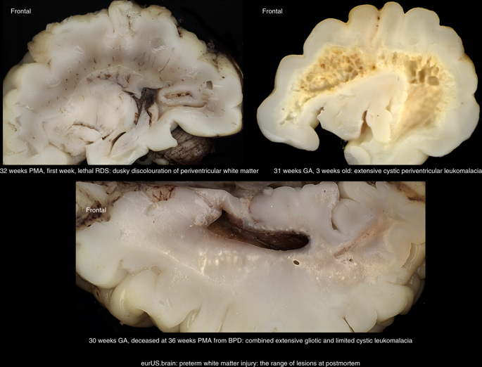
White matter injury (WMI) is the most frequent form of preterm brain injury. Cranial ultrasound (CUS) remains the preferred modality for initial and sequential neuroimaging in preterm infants, and is reliable for the diagnosis of cystic periventricular leukomalacia. Although magnetic resonance imaging is superior to CUS in detecting the diffuse and more subtle forms of WMI that prevail in very premature infants surviving nowadays, recent improvement in the quality of neonatal CUS imaging has broadened the spectrum of preterm white matter abnormalities that can be detected with this technique. We propose a structured CUS assessment of WMI of prematurity that seeks to account for both cystic and non-cystic changes, as well as signs of white matter loss and impaired brain growth and maturation, at or near term equivalent age. This novel assessment system aims to improve disease description in both routine clinical practice and clinical research. Whether this systematic assessment will improve prediction of outcome in preterm infants with WMI still needs to be evaluated in prospective studies.
中文翻译:

早产白质损伤:超声诊断和分类
白质损伤 (WMI) 是最常见的早产脑损伤形式。颅脑超声 (CUS) 仍然是早产儿初始和顺序神经影像学检查的首选方式,对于囊性脑室周围白质软化症的诊断是可靠的。尽管磁共振成像在检测目前幸存的极早产儿中普遍存在的弥漫性和更细微的 WMI 形式方面优于 CUS,但最近新生儿 CUS 成像质量的提高拓宽了可检测到的早产白质异常的范围。这种技术。我们建议对早产 WMI 进行结构化的 CUS 评估,旨在解释囊性和非囊性变化,以及白质丢失和大脑生长和成熟受损的迹象,在或接近等效年龄。这种新颖的评估系统旨在改进常规临床实践和临床研究中的疾病描述。这种系统评估是否会改善对 WMI 早产儿预后的预测,仍需要在前瞻性研究中进行评估。
更新日期:2020-03-01
中文翻译:
早产白质损伤:超声诊断和分类
白质损伤 (WMI) 是最常见的早产脑损伤形式。颅脑超声 (CUS) 仍然是早产儿初始和顺序神经影像学检查的首选方式,对于囊性脑室周围白质软化症的诊断是可靠的。尽管磁共振成像在检测目前幸存的极早产儿中普遍存在的弥漫性和更细微的 WMI 形式方面优于 CUS,但最近新生儿 CUS 成像质量的提高拓宽了可检测到的早产白质异常的范围。这种技术。我们建议对早产 WMI 进行结构化的 CUS 评估,旨在解释囊性和非囊性变化,以及白质丢失和大脑生长和成熟受损的迹象,在或接近等效年龄。这种新颖的评估系统旨在改进常规临床实践和临床研究中的疾病描述。这种系统评估是否会改善对 WMI 早产儿预后的预测,仍需要在前瞻性研究中进行评估。


 京公网安备 11010802027423号