当前位置:
X-MOL 学术
›
PLOS Pathog.
›
论文详情
Our official English website, www.x-mol.net, welcomes your
feedback! (Note: you will need to create a separate account there.)
Murine polyomavirus DNA transitions through spatially distinct nuclear replication subdomains during infection.
PLoS Pathogens ( IF 5.5 ) Pub Date : 2020-03-23 , DOI: 10.1371/journal.ppat.1008403 Douglas K Peters 1 , Robert L Garcea 1, 2
PLoS Pathogens ( IF 5.5 ) Pub Date : 2020-03-23 , DOI: 10.1371/journal.ppat.1008403 Douglas K Peters 1 , Robert L Garcea 1, 2
Affiliation
|
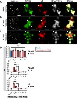
The replication of small DNA viruses requires both host DNA replication and repair factors that are often recruited to subnuclear domains termed viral replication centers (VRCs). Aside from serving as a spatial focus for viral replication, little is known about these dynamic areas in the nucleus. We investigated the organization and function of VRCs during murine polyomavirus (MuPyV) infection using 3D structured illumination microscopy (3D-SIM). We localized MuPyV replication center components, such as the viral large T-antigen (LT) and the cellular replication protein A (RPA), to spatially distinct subdomains within VRCs. We found that viral DNA (vDNA) trafficked sequentially through these subdomains post-synthesis, suggesting their distinct functional roles in vDNA processing. Additionally, we observed disruption of VRC organization and vDNA trafficking during mutant MuPyV infections or inhibition of DNA synthesis. These results reveal a dynamic organization of VRC components that coordinates virus replication.
中文翻译:

鼠多瘤病毒 DNA 在感染过程中通过空间上不同的核复制子域进行转变。
小 DNA 病毒的复制需要宿主 DNA 复制和修复因子,这些因子通常被招募到称为病毒复制中心 (VRC) 的亚核域。除了作为病毒复制的空间焦点之外,人们对细胞核中的这些动态区域知之甚少。我们使用 3D 结构照明显微镜 (3D-SIM) 研究了鼠多瘤病毒 (MuPyV) 感染期间 VRC 的组织和功能。我们将 MuPyV 复制中心组件,例如病毒大 T 抗原 (LT) 和细胞复制蛋白 A (RPA) 定位到 VRC 内空间上不同的子域。我们发现病毒 DNA (vDNA) 在合成后依次通过这些子域,表明它们在 vDNA 加工中具有独特的功能作用。此外,我们观察到突变 MuPyV 感染期间 VRC 组织和 vDNA 运输的破坏或 DNA 合成的抑制。这些结果揭示了协调病毒复制的 VRC 组件的动态组织。
更新日期:2020-03-24
中文翻译:
鼠多瘤病毒 DNA 在感染过程中通过空间上不同的核复制子域进行转变。
小 DNA 病毒的复制需要宿主 DNA 复制和修复因子,这些因子通常被招募到称为病毒复制中心 (VRC) 的亚核域。除了作为病毒复制的空间焦点之外,人们对细胞核中的这些动态区域知之甚少。我们使用 3D 结构照明显微镜 (3D-SIM) 研究了鼠多瘤病毒 (MuPyV) 感染期间 VRC 的组织和功能。我们将 MuPyV 复制中心组件,例如病毒大 T 抗原 (LT) 和细胞复制蛋白 A (RPA) 定位到 VRC 内空间上不同的子域。我们发现病毒 DNA (vDNA) 在合成后依次通过这些子域,表明它们在 vDNA 加工中具有独特的功能作用。此外,我们观察到突变 MuPyV 感染期间 VRC 组织和 vDNA 运输的破坏或 DNA 合成的抑制。这些结果揭示了协调病毒复制的 VRC 组件的动态组织。


 京公网安备 11010802027423号