当前位置:
X-MOL 学术
›
Anal. Chem.
›
论文详情
Our official English website, www.x-mol.net, welcomes your
feedback! (Note: you will need to create a separate account there.)
Multimodal Multiphoton Imaging of the Lipid Bilayer by Dye-Based Sum-Frequency Generation and Coherent Anti-Stokes Raman Scattering.
Analytical Chemistry ( IF 6.7 ) Pub Date : 2020-03-27 , DOI: 10.1021/acs.analchem.0c00673 Takaha Mizuguchi 1, 2 , Atsuya Momotake 3 , Mafumi Hishida 3 , Masato Yasui 1, 4 , Yasuhiko Yamamoto 3 , Toshiharu Saiki 2 , Mutsuo Nuriya 1, 4, 5, 6
Analytical Chemistry ( IF 6.7 ) Pub Date : 2020-03-27 , DOI: 10.1021/acs.analchem.0c00673 Takaha Mizuguchi 1, 2 , Atsuya Momotake 3 , Mafumi Hishida 3 , Masato Yasui 1, 4 , Yasuhiko Yamamoto 3 , Toshiharu Saiki 2 , Mutsuo Nuriya 1, 4, 5, 6
Affiliation
|
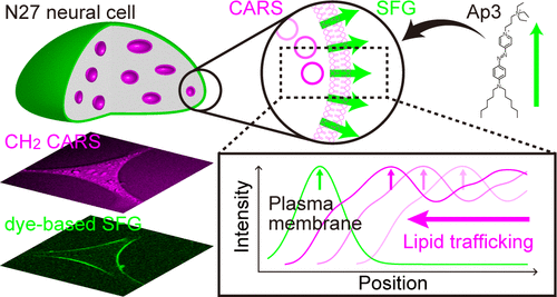
Coherent anti-Stokes Raman scattering (CARS) imaging is widely used for imaging molecular vibrations inside cells and tissues. Lipid bilayers are potential analytes for CARS imaging due to their abundant CH2 vibrational bonds. However, identifying the plasma membrane is challenging since it possesses a thin structure and is closely apposed to lipid structures inside the cells. Since the plasma membrane provides the most prominent asymmetric location within cells, orientation sensitive sum-frequency generation (SFG) imaging is a promising technique for selective visualization of the plasma membrane labeled by a nonfluorescent and SFG-specific dye, Ap3, when using a CARS microscope system. In this study, we closely compare the characteristics of lipid bilayer imaging by dye-based SFG and CARS using giant vesicles (GVs) and N27 rat dopaminergic neural cells. As a result, we show that CARS imaging can be exploited for the visualization of whole lipid structures inside GVs and cells but is insufficient for identification of the plasma membrane, which instead can be achieved using dye-based SFG imaging. In addition, we demonstrate that these unique properties can be combined and applied to the live-cell tracking of intracellular lipid structures such as lipid droplets beneath the plasma membrane. Thus, multimodal multiphoton imaging through a combination of dye-based SFG and CARS can serve as a powerful chemical imaging tool to investigate lipid bilayers in GVs and living cells.
中文翻译:

通过基于染料的总和生成和相干抗斯托克斯拉曼散射对脂质双层的多峰多光子成像。
相干抗斯托克斯拉曼散射(CARS)成像被广泛用于成像细胞和组织内部的分子振动。脂质双层由于其丰富的CH2振动键而成为CARS成像的潜在分析物。然而,鉴定质膜是具有挑战性的,因为它具有薄的结构并且与细胞内部的脂质结构紧密相关。由于质膜在细胞内提供了最突出的不对称位置,因此在使用CARS时,定向敏感的和频生成(SFG)成像是一种选择性可视化非荧光和SFG特定染料Ap3标记的质膜的有前途的技术显微镜系统。在这个研究中,我们使用巨囊泡(GV)和N27大鼠多巴胺能神经细胞通过染料基SFG和CARS紧密比较脂质双层成像的特征。结果,我们表明CARS成像可用于可视化GV和细胞内部的完整脂质结构,但不足以识别质膜,而可以使用基于染料的SFG成像来实现。此外,我们证明了这些独特的特性可以组合起来,并应用于活体内细胞内脂质结构的追踪,例如质膜下的脂质滴。因此,通过基于染料的SFG和CARS的组合进行的多峰多光子成像可以用作研究GV和活细胞中脂质双层的强大化学成像工具。我们显示,CARS成像可用于可视化GV和细胞内的整个脂质结构,但不足以鉴定质膜,而可以使用基于染料的SFG成像来实现。此外,我们证明了这些独特的特性可以组合起来,并应用于活体内细胞内脂质结构的追踪,例如质膜下的脂质滴。因此,通过基于染料的SFG和CARS的组合进行的多峰多光子成像可以用作研究GV和活细胞中脂质双层的强大化学成像工具。我们显示,CARS成像可用于可视化GV和细胞内的整个脂质结构,但不足以鉴定质膜,而可以使用基于染料的SFG成像来实现。此外,我们证明了这些独特的特性可以组合起来,并应用于活体内细胞内脂质结构的追踪,例如质膜下的脂质滴。因此,通过基于染料的SFG和CARS的组合进行的多峰多光子成像可以用作研究GV和活细胞中脂质双层的强大化学成像工具。我们证明了这些独特的特性可以组合起来,并应用于细胞内脂质结构(例如质膜下的脂质滴)的活细胞追踪。因此,通过基于染料的SFG和CARS的组合进行的多峰多光子成像可以用作研究GV和活细胞中脂质双层的强大化学成像工具。我们证明了这些独特的特性可以组合起来,并应用于细胞内脂质结构(例如质膜下的脂质滴)的活细胞跟踪。因此,通过基于染料的SFG和CARS的组合进行的多峰多光子成像可以用作研究GV和活细胞中脂质双层的强大化学成像工具。
更新日期:2020-04-23
中文翻译:
通过基于染料的总和生成和相干抗斯托克斯拉曼散射对脂质双层的多峰多光子成像。
相干抗斯托克斯拉曼散射(CARS)成像被广泛用于成像细胞和组织内部的分子振动。脂质双层由于其丰富的CH2振动键而成为CARS成像的潜在分析物。然而,鉴定质膜是具有挑战性的,因为它具有薄的结构并且与细胞内部的脂质结构紧密相关。由于质膜在细胞内提供了最突出的不对称位置,因此在使用CARS时,定向敏感的和频生成(SFG)成像是一种选择性可视化非荧光和SFG特定染料Ap3标记的质膜的有前途的技术显微镜系统。在这个研究中,我们使用巨囊泡(GV)和N27大鼠多巴胺能神经细胞通过染料基SFG和CARS紧密比较脂质双层成像的特征。结果,我们表明CARS成像可用于可视化GV和细胞内部的完整脂质结构,但不足以识别质膜,而可以使用基于染料的SFG成像来实现。此外,我们证明了这些独特的特性可以组合起来,并应用于活体内细胞内脂质结构的追踪,例如质膜下的脂质滴。因此,通过基于染料的SFG和CARS的组合进行的多峰多光子成像可以用作研究GV和活细胞中脂质双层的强大化学成像工具。我们显示,CARS成像可用于可视化GV和细胞内的整个脂质结构,但不足以鉴定质膜,而可以使用基于染料的SFG成像来实现。此外,我们证明了这些独特的特性可以组合起来,并应用于活体内细胞内脂质结构的追踪,例如质膜下的脂质滴。因此,通过基于染料的SFG和CARS的组合进行的多峰多光子成像可以用作研究GV和活细胞中脂质双层的强大化学成像工具。我们显示,CARS成像可用于可视化GV和细胞内的整个脂质结构,但不足以鉴定质膜,而可以使用基于染料的SFG成像来实现。此外,我们证明了这些独特的特性可以组合起来,并应用于活体内细胞内脂质结构的追踪,例如质膜下的脂质滴。因此,通过基于染料的SFG和CARS的组合进行的多峰多光子成像可以用作研究GV和活细胞中脂质双层的强大化学成像工具。我们证明了这些独特的特性可以组合起来,并应用于细胞内脂质结构(例如质膜下的脂质滴)的活细胞追踪。因此,通过基于染料的SFG和CARS的组合进行的多峰多光子成像可以用作研究GV和活细胞中脂质双层的强大化学成像工具。我们证明了这些独特的特性可以组合起来,并应用于细胞内脂质结构(例如质膜下的脂质滴)的活细胞跟踪。因此,通过基于染料的SFG和CARS的组合进行的多峰多光子成像可以用作研究GV和活细胞中脂质双层的强大化学成像工具。


 京公网安备 11010802027423号