当前位置:
X-MOL 学术
›
Exp. Eye Res.
›
论文详情
Our official English website, www.x-mol.net, welcomes your
feedback! (Note: you will need to create a separate account there.)
Axonal debris accumulates in corneal epithelial cells after intraepithelial corneal nerves are damaged: A focused Ion Beam Scanning Electron Microscopy (FIB-SEM) study.
Experimental Eye Research ( IF 3.0 ) Pub Date : 2020-03-21 , DOI: 10.1016/j.exer.2020.107998 Paola Parlanti 1 , Sonali Pal-Ghosh 2 , Alexa Williams 2 , Gauri Tadvalkar 2 , Anastas Popratiloff 3 , Mary Ann Stepp 4
Experimental Eye Research ( IF 3.0 ) Pub Date : 2020-03-21 , DOI: 10.1016/j.exer.2020.107998 Paola Parlanti 1 , Sonali Pal-Ghosh 2 , Alexa Williams 2 , Gauri Tadvalkar 2 , Anastas Popratiloff 3 , Mary Ann Stepp 4
Affiliation
|
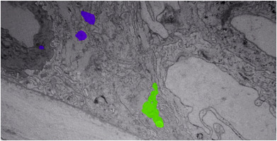
The intraepithelial corneal nerves (ICNs) that innervate the corneal epithelium are maintained through interactions with corneal epithelial cells and the extracellular matrix they produce. One to several axons bundle together within the basal cell layer and extend parallel to the ocular surface or branch and extend apically. Here we use 3-dimentional (3D) ultrastructural reconstructions of control and trephine injured mouse corneal epithelium and stroma produced using Focused Ion Beam Scanning Electron Microscope (FIB-SEM) to determine whether corneal epithelial or immune cells resident in the epithelium remove axonal debris and degrade it in their lysosomes after trephine injury to the cornea. We demonstrate that axonal fragments are internalized in the corneal epithelium and accumulate within electron dense structures consistent with lysosomes 3 h after trephine injury in both epithelial and immune cells located among the basal cells of the trephine injured cornea. Confocal imaging showed fewer CD45+ immune cells within the corneal epithelium after trephine injury compared to controls. The resolution obtained using FIB-SEM also allowed us to show that the presence of sensory axons at the basal aspect of the epithelial basal cells close to the anterior aspect of the epithelial basement membrane (EBM) is associated with a focal reduction in EBM thickness. In addition, we show using FIB-SEM and confocal imaging that superficial trephine injuries that do not penetrate the stroma, damage the integrity of anterior stromal nerves. These studies are the first to look at the mouse cornea following nerve injury using FIB-SEM.
中文翻译:

上皮内角膜神经受损后,轴突碎片积聚在角膜上皮细胞中:聚焦离子束扫描电子显微镜 (FIB-SEM) 研究。
支配角膜上皮的上皮内角膜神经 (ICN) 通过与角膜上皮细胞及其产生的细胞外基质的相互作用来维持。一到几个轴突在基底细胞层内捆绑在一起,平行于眼表或分支延伸并延伸到顶端。在这里,我们使用聚焦离子束扫描电子显微镜 (FIB-SEM) 产生的对照和环钻损伤的小鼠角膜上皮和基质的 3 维 (3D) 超微结构重建来确定角膜上皮或免疫细胞是否位于上皮中去除轴突碎片和环钻损伤角膜后,在溶酶体中降解它。我们证明轴突片段在角膜上皮中内化,并在环钻损伤后 3 小时在位于环钻损伤角膜的基底细胞之间的上皮细胞和免疫细胞中积累在与溶酶体一致的电子致密结构内。与对照组相比,共聚焦成像显示环钻损伤后角膜上皮内的 CD45+ 免疫细胞较少。使用 FIB-SEM 获得的分辨率还使我们能够表明,在靠近上皮基底膜 (EBM) 前部的上皮基底细胞基底面存在感觉轴突与 EBM 厚度的局部减少有关。此外,我们使用 FIB-SEM 和共聚焦成像显示,不穿透间质的浅表环钻损伤会损害前间质神经的完整性。
更新日期:2020-03-22
中文翻译:
上皮内角膜神经受损后,轴突碎片积聚在角膜上皮细胞中:聚焦离子束扫描电子显微镜 (FIB-SEM) 研究。
支配角膜上皮的上皮内角膜神经 (ICN) 通过与角膜上皮细胞及其产生的细胞外基质的相互作用来维持。一到几个轴突在基底细胞层内捆绑在一起,平行于眼表或分支延伸并延伸到顶端。在这里,我们使用聚焦离子束扫描电子显微镜 (FIB-SEM) 产生的对照和环钻损伤的小鼠角膜上皮和基质的 3 维 (3D) 超微结构重建来确定角膜上皮或免疫细胞是否位于上皮中去除轴突碎片和环钻损伤角膜后,在溶酶体中降解它。我们证明轴突片段在角膜上皮中内化,并在环钻损伤后 3 小时在位于环钻损伤角膜的基底细胞之间的上皮细胞和免疫细胞中积累在与溶酶体一致的电子致密结构内。与对照组相比,共聚焦成像显示环钻损伤后角膜上皮内的 CD45+ 免疫细胞较少。使用 FIB-SEM 获得的分辨率还使我们能够表明,在靠近上皮基底膜 (EBM) 前部的上皮基底细胞基底面存在感觉轴突与 EBM 厚度的局部减少有关。此外,我们使用 FIB-SEM 和共聚焦成像显示,不穿透间质的浅表环钻损伤会损害前间质神经的完整性。


 京公网安备 11010802027423号