Biomaterials Advances ( IF 5.5 ) Pub Date : 2020-03-21 , DOI: 10.1016/j.msec.2020.110860 Karan Gulati 1 , Ho-Jin Moon 2 , P T Sudheesh Kumar 3 , Pingping Han 1 , Sašo Ivanovski 1
|
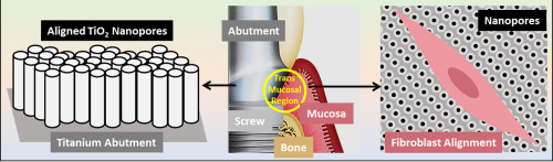
Ensuring the formation of a robust trans-mucosal soft-tissue seal at the dental abutment surface is crucial towards protecting the underlying dental implant associated tissues from the external microbial-rich oral environment. The ability to mechanically enhance fibroblast functions at the dental abutment-mucosa interface, without the use of bioactive agents, holds great promise towards reducing the ingress of oral pathogens into the dental implant microenvironment. We hereby propose fabrication of unique anisotropic titania nanopores (TNPs) on the surface of titanium (via electrochemical anodization, EA) towards enhancing the soft-tissue integration and wound healing abilities of the conventional abutments. Using optimized EA, mechanically robust TNPs of varied diameters were fabricated on Ti surfaces with preserved underlying substrate micro-features: dual micro-nanostructured surfaces. Next, we evaluated the mechanical stability of such structures and demonstrated the ease of fabrication on commercial abutment geometries. The functions of primary human gingival fibroblasts (GFs) cultured on these surfaces in vitro were evaluated from 1 h to 7 days, and were compared between TNPs and clinically relevant titanium controls: as-received irregular rough Ti (Rough Ti) and mechanically prepared micro-rough Ti (Micro Ti). Improved cell viability was observed on TNPs as compared to controls. Additionally, cellular spreading morphology indicated cell alignment along the direction of the nanopores with strong anchoring evident by enhanced filopodia and stress fibers. RT-PCR showed improved wound healing, cell migration/adhesion and angiogenesis related mRNA, especially for TNPs with large diameters. This study provides a proof-of-concept towards using anodization for improving soft-tissue sealing around dental abutment surfaces, with implications towards reducing implant failure/peri-implantitis and achieving long-term success, especially in compromised patient conditions.
中文翻译:
阳极氧化的各向异性钛表面可增强对牙龈成纤维细胞的引导。
确保在牙齿基台表面形成牢固的跨粘膜软组织密封对于保护下面的与牙齿植入物相关的组织免受外部富含微生物的口腔环境的影响至关重要。在不使用生物活性剂的情况下,机械增强牙齿基台-粘膜界面上的成纤维细胞功能的能力,有望减少口腔病原体进入牙齿植入物微环境。我们据此提出在钛表面上制备独特的各向异性二氧化钛纳米孔(TNP)(通过电化学阳极氧化,EA),以增强传统基台的软组织整合和伤口愈合能力。使用优化的EA,在具有保留的底层基底微特征的Ti表面上制造了直径可变的机械坚固的TNP:双重微纳米结构表面。接下来,我们评估了此类结构的机械稳定性,并证明了在商业基台几何形状上制造的简便性。在这些表面上培养的原代人牙龈成纤维细胞(GFs)的功能体外从1小时到7天进行了评估,并在TNP和临床相关的钛对照之间进行了比较:按原样接收的不规则粗Ti(Rough Ti)和机械制备的微粗Ti(Micro Ti)。与对照相比,在TNP上观察到细胞活力的提高。另外,细胞扩散形态表明细胞沿着纳米孔的方向排列,通过增强的丝状伪足和应激纤维表现出强烈的锚定作用。RT-PCR显示改善的伤口愈合,细胞迁移/粘附和血管生成相关的mRNA,特别是对于大直径的TNP。这项研究为使用阳极氧化技术改善牙基台表面周围的软组织密封提供了概念验证,并有助于减少种植体失败/种植体周围炎并取得长期成功,


 京公网安备 11010802027423号