当前位置:
X-MOL 学术
›
Anal. Chem.
›
论文详情
Our official English website, www.x-mol.net, welcomes your
feedback! (Note: you will need to create a separate account there.)
In Vivo Imaging of Hypoxia Associated with Inflammatory Bowel Disease by a Cytoplasmic Protein-Powered Fluorescence Cascade Amplifier.
Analytical Chemistry ( IF 6.7 ) Pub Date : 2020-03-31 , DOI: 10.1021/acs.analchem.9b05278 Yibo Zhou 1 , Sheng Yang 1 , Jingru Guo 1 , Hao Dong 1 , Keyi Yin 1 , Wei Tao Huang 2 , Ronghua Yang 1, 3
Analytical Chemistry ( IF 6.7 ) Pub Date : 2020-03-31 , DOI: 10.1021/acs.analchem.9b05278 Yibo Zhou 1 , Sheng Yang 1 , Jingru Guo 1 , Hao Dong 1 , Keyi Yin 1 , Wei Tao Huang 2 , Ronghua Yang 1, 3
Affiliation
|
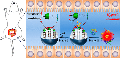
Accurate and sensitive imaging of hypoxia associated with inflammatory bowel disease (IBD) is significant for the precise diagnosis and treatment of this disease, but it remains a challenge for traditional hypoxia-activatable fluorescence probes because of a more moderate hypoxic state during IBD than under other pathological conditions. To address this issue, herein, we designed a hypoxia-activatable and cytoplasmic protein-powered fluorescence cascade amplifier, named HCFA, to image hypoxia associated with IBD in vivo. In our design, a 4-aminobenzoic acid (azo)-modified mesoporous silica nanoparticle (MSN) was used as a container to load black hole quencher 2 (BHQ2) and cytoplasmic protein-binding squarylium dye (SQ); then, the β-cyclodextrin polymer (β-CDP) combined with azo through a host-guest interaction to form HCFA. Upon passive stagnation in the inflamed tissue of IBD, the azo band would be cleaved under a hypoxic microenvironment, and SQ was released to activate the fluorescence of HCFA. Moreover, the unconstrained SQ can bind with cytoplasmic protein to exhibit drastic fluorescence intensity enhancement, realizing the fluorescence signal amplification for imaging of hypoxia. When one takes advantage of the large load capacity of MSN and the unique property of SQ, HCFA can sense oxygen levels in the range of 0% to 10%. Meanwhile, the fluorescence imaging results demonstrate that HCFA can sensitively distinguish different levels of cellular hypoxia and monitor the variations of hypoxia in vivo, highlighting HCFA as a promising tool for the detection of hypoxia associated with IBD.
中文翻译:

通过细胞质蛋白供电的荧光级联放大器对与炎症性肠病相关的缺氧的体内成像。
与炎症性肠病(IBD)相关的低氧的准确而敏感的成像对于精确诊断和治疗该疾病具有重要意义,但是由于IBD期间的低氧状态比其他低氧激活的荧光探针更为温和,因此对于传统的可低氧激活的荧光探针仍然是一个挑战。病理状况。为了解决这个问题,在本文中,我们设计了一种名为HCFA的低氧激活型和细胞质蛋白供电的荧光级联放大器,以在体内成像与IBD相关的低氧。在我们的设计中,使用4-氨基苯甲酸(偶氮)改性的介孔二氧化硅纳米粒子(MSN)作为容器,以加载黑洞淬灭剂2(BHQ2)和胞质蛋白结合方酸染料(SQ)。然后,β-环糊精聚合物(β-CDP)通过主体-客体相互作用与偶氮结合形成HCFA。在IBD发炎组织中被动停滞后,在低氧微环境下偶氮带会断裂,SQ被释放以激活HCFA的荧光。此外,不受约束的SQ可以与细胞质蛋白结合,表现出急剧的荧光强度增强,实现了对缺氧成像的荧光信号放大。当人们利用MSN的大负载能力和SQ的独特性能时,HCFA可以感应到0%至10%的氧气含量。同时,荧光成像结果表明,HCFA可以灵敏地区分不同水平的细胞缺氧并监测体内缺氧的变化,突出显示HCFA是检测与IBD相关的缺氧的有前途的工具。在缺氧的微环境下,偶氮带会断裂,SQ被释放以激活HCFA的荧光。此外,不受约束的SQ可以与细胞质蛋白结合,表现出急剧的荧光强度增强,实现了对缺氧成像的荧光信号放大。当人们利用MSN的大负载能力和SQ的独特性能时,HCFA可以感应到0%至10%的氧气含量。同时,荧光成像结果表明,HCFA可以灵敏地区分不同水平的细胞缺氧并监测体内缺氧的变化,突出显示HCFA是检测与IBD相关的缺氧的有前途的工具。在缺氧的微环境下,偶氮带会断裂,SQ被释放以激活HCFA的荧光。此外,不受约束的SQ可以与细胞质蛋白结合,表现出急剧的荧光强度增强,实现了对缺氧成像的荧光信号放大。当人们利用MSN的大负载能力和SQ的独特性能时,HCFA可以感应到0%至10%的氧气含量。同时,荧光成像结果表明,HCFA可以灵敏地区分不同水平的细胞缺氧并监测体内缺氧的变化,突出显示HCFA是检测与IBD相关的缺氧的有前途的工具。不受约束的SQ可以与细胞质蛋白结合,表现出急剧的荧光强度增强,实现了对缺氧成像的荧光信号放大。当人们利用MSN的大负载能力和SQ的独特性能时,HCFA可以感应到0%至10%的氧气含量。同时,荧光成像结果表明,HCFA可以灵敏地区分不同水平的细胞缺氧并监测体内缺氧的变化,突出显示HCFA是检测与IBD相关的缺氧的有前途的工具。不受约束的SQ可以与细胞质蛋白结合,表现出急剧的荧光强度增强,实现了对缺氧成像的荧光信号放大。当人们利用MSN的大负载能力和SQ的独特性能时,HCFA可以感应到0%至10%的氧气含量。同时,荧光成像结果表明,HCFA可以敏感地区分不同水平的细胞缺氧并监测体内缺氧的变化,突出显示HCFA是检测与IBD相关的缺氧的有前途的工具。HCFA可以检测到0%至10%的氧气含量。同时,荧光成像结果表明,HCFA可以灵敏地区分不同水平的细胞缺氧并监测体内缺氧的变化,突出显示HCFA是检测与IBD相关的缺氧的有前途的工具。HCFA可以检测到0%至10%的氧气含量。同时,荧光成像结果表明,HCFA可以敏感地区分不同水平的细胞缺氧并监测体内缺氧的变化,突出显示HCFA是检测与IBD相关的缺氧的有前途的工具。
更新日期:2020-04-23
中文翻译:
通过细胞质蛋白供电的荧光级联放大器对与炎症性肠病相关的缺氧的体内成像。
与炎症性肠病(IBD)相关的低氧的准确而敏感的成像对于精确诊断和治疗该疾病具有重要意义,但是由于IBD期间的低氧状态比其他低氧激活的荧光探针更为温和,因此对于传统的可低氧激活的荧光探针仍然是一个挑战。病理状况。为了解决这个问题,在本文中,我们设计了一种名为HCFA的低氧激活型和细胞质蛋白供电的荧光级联放大器,以在体内成像与IBD相关的低氧。在我们的设计中,使用4-氨基苯甲酸(偶氮)改性的介孔二氧化硅纳米粒子(MSN)作为容器,以加载黑洞淬灭剂2(BHQ2)和胞质蛋白结合方酸染料(SQ)。然后,β-环糊精聚合物(β-CDP)通过主体-客体相互作用与偶氮结合形成HCFA。在IBD发炎组织中被动停滞后,在低氧微环境下偶氮带会断裂,SQ被释放以激活HCFA的荧光。此外,不受约束的SQ可以与细胞质蛋白结合,表现出急剧的荧光强度增强,实现了对缺氧成像的荧光信号放大。当人们利用MSN的大负载能力和SQ的独特性能时,HCFA可以感应到0%至10%的氧气含量。同时,荧光成像结果表明,HCFA可以灵敏地区分不同水平的细胞缺氧并监测体内缺氧的变化,突出显示HCFA是检测与IBD相关的缺氧的有前途的工具。在缺氧的微环境下,偶氮带会断裂,SQ被释放以激活HCFA的荧光。此外,不受约束的SQ可以与细胞质蛋白结合,表现出急剧的荧光强度增强,实现了对缺氧成像的荧光信号放大。当人们利用MSN的大负载能力和SQ的独特性能时,HCFA可以感应到0%至10%的氧气含量。同时,荧光成像结果表明,HCFA可以灵敏地区分不同水平的细胞缺氧并监测体内缺氧的变化,突出显示HCFA是检测与IBD相关的缺氧的有前途的工具。在缺氧的微环境下,偶氮带会断裂,SQ被释放以激活HCFA的荧光。此外,不受约束的SQ可以与细胞质蛋白结合,表现出急剧的荧光强度增强,实现了对缺氧成像的荧光信号放大。当人们利用MSN的大负载能力和SQ的独特性能时,HCFA可以感应到0%至10%的氧气含量。同时,荧光成像结果表明,HCFA可以灵敏地区分不同水平的细胞缺氧并监测体内缺氧的变化,突出显示HCFA是检测与IBD相关的缺氧的有前途的工具。不受约束的SQ可以与细胞质蛋白结合,表现出急剧的荧光强度增强,实现了对缺氧成像的荧光信号放大。当人们利用MSN的大负载能力和SQ的独特性能时,HCFA可以感应到0%至10%的氧气含量。同时,荧光成像结果表明,HCFA可以灵敏地区分不同水平的细胞缺氧并监测体内缺氧的变化,突出显示HCFA是检测与IBD相关的缺氧的有前途的工具。不受约束的SQ可以与细胞质蛋白结合,表现出急剧的荧光强度增强,实现了对缺氧成像的荧光信号放大。当人们利用MSN的大负载能力和SQ的独特性能时,HCFA可以感应到0%至10%的氧气含量。同时,荧光成像结果表明,HCFA可以敏感地区分不同水平的细胞缺氧并监测体内缺氧的变化,突出显示HCFA是检测与IBD相关的缺氧的有前途的工具。HCFA可以检测到0%至10%的氧气含量。同时,荧光成像结果表明,HCFA可以灵敏地区分不同水平的细胞缺氧并监测体内缺氧的变化,突出显示HCFA是检测与IBD相关的缺氧的有前途的工具。HCFA可以检测到0%至10%的氧气含量。同时,荧光成像结果表明,HCFA可以敏感地区分不同水平的细胞缺氧并监测体内缺氧的变化,突出显示HCFA是检测与IBD相关的缺氧的有前途的工具。


 京公网安备 11010802027423号