Spinal Cord ( IF 2.2 ) Pub Date : 2020-03-19 , DOI: 10.1038/s41393-020-0442-6 Nao Tamai 1, 2 , Takeo Minematsu 2, 3 , Tomonori Maeda 4 , Koichi Yabunaka 1, 2 , Hiromi Sanada 2, 4
|
Study design
Secondary analysis of a cross-sectional observation study.
Objectives
To determine the relationship between skin ultrasound images and muscle damage in wheelchair basketball athletes, using skin blotting examinations of the ischial regions.
Setting
Community, Japan.
Methods
Fourteen elite wheelchair basketball athletes were recruited. We obtained data regarding participants’ characteristics. We undertook ultrasonographic images and quantitative skin blotting of the ischial region before and after training, and after rest.
Results
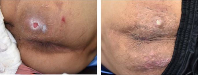
We identified Category II and III pressure injuries in 2 of the 12 participants. Structural features were classified into four categories based on ultrasonographic features, namely, normal skin structure, unclear superficial and deep fascia, cloudy fat layer, and fat infiltration and low-echoic lesion/anechoic lesions. The muscle-type creatinine kinase (CK-M) level (median [interquartile range: IQR], 2.98 [2.80–3.47]) in the fat infiltration and low-echoic lesion/anechoic lesion group was significantly higher (1.43 [1.41–1.49]) than in a nonfat infiltration and low-echoic lesion/anechoic lesion group after training (p = 0.03). The interleukin-6 (IL-6) level (median [IQR], 23.5 [16.15–58.97]) in the fat infiltration and low-echoic lesion/anechoic lesion group was significantly higher (1.94 [1.74–4.44]) than in the nonfat infiltration and low-echoic lesion/anechoic lesion group after rest (mean difference = −25.4, 95% CI −61.1 to 10.7, p = 0.03).
Conclusions
The combination of ultrasonographic images and skin blotting using CK-M and IL-6, could detect early deep tissue damage in wheelchair athletes. These techniques could be potentially useful in the treatment and prevention of pressure injuries.
Sponsorship
This study was supported in part by YAMAHA Motor Foundation for Sports.
中文翻译:
轮椅篮球运动员中皮肤超声图像与使用皮肤印迹的肌肉损伤之间的关系。
学习规划
横截面观察研究的二级分析。
目标
使用坐骨区域的皮肤印迹检查来确定轮椅篮球运动员皮肤超声图像与肌肉损伤之间的关系。
设置
日本社区。
方法
招募了14名精英轮椅篮球运动员。我们获得了有关参与者特征的数据。我们在训练前后,休息后进行了坐骨区域的超声图像和定量皮肤印迹分析。
结果
我们在12名参与者中的2名中确定了II类和III类压力损伤。根据超声特征,将结构特征分为四类,即正常皮肤结构,浅浅和深筋膜不清楚,脂肪层浑浊,脂肪浸润和低回声病变/无回声病变。脂肪浸润和低回声病变/无回声病变组的肌肉型肌酐激酶(CK-M)水平(中位[四分位数范围:IQR],2.98 [2.80–3.47])显着更高(1.43 [1.41–1.49] ])比训练后的非脂肪浸润和低回声病变/无回声病变组(p = 0.03)。脂肪浸润和低回声病变/无回声病变组的白细胞介素6(IL-6)水平(中位值[IQR],23.5 [16.15–58.97])显着高于白细胞介素6(IL-4)水平(1.94 [1.74–4.44])休息后脱脂浸润和低回声病变/无回声病变组(平均差异= -25.4,95%CI -61.1至10.7,p = 0.03)。
结论
超声图像和使用CK-M和IL-6的皮肤印迹相结合,可以检测轮椅运动员早期的深部组织损伤。这些技术可能在治疗和预防压力损伤中可能有用。
赞助商
这项研究得到了YAMAHA运动体育基金会的部分支持。


 京公网安备 11010802027423号