当前位置:
X-MOL 学术
›
Colloids Surf. B Biointerfaces
›
论文详情
Our official English website, www.x-mol.net, welcomes your feedback! (Note: you will need to create a separate account there.)
Regulated phase separation in nanopatterned protein-polysaccharide thin films by spin coating.
Colloids and Surfaces B: Biointerfaces ( IF 5.8 ) Pub Date : 2020-03-18 , DOI: 10.1016/j.colsurfb.2020.110967 Russell A Banta 1 , Timothy W Collins 1 , Ricky Curley 1 , John O'Connell 1 , Paul W Young 2 , Justin D Holmes 1 , Eoin J Flynn 1
Colloids and Surfaces B: Biointerfaces ( IF 5.8 ) Pub Date : 2020-03-18 , DOI: 10.1016/j.colsurfb.2020.110967 Russell A Banta 1 , Timothy W Collins 1 , Ricky Curley 1 , John O'Connell 1 , Paul W Young 2 , Justin D Holmes 1 , Eoin J Flynn 1
Affiliation
|
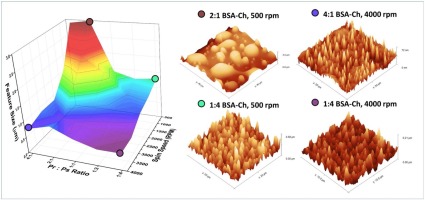
Patterned films are essential to the commonplace technologies of modern life. However, they come at high cost to the planet, being produced from non-renewable, petrochemical-derived polymers and utilising substrates that require harsh, top-down etching techniques. Biopolymers offer a cheap, sustainable and viable alternative easily integrated into existing production techniques. We describe a simple method for the production of patterned biopolymer surfaces and the assignment of each biopolymer domain, which allows for selective metal incorporation used in many patterning applications. Protein and polysaccharide domains were identified by selective etching and metal incorporation; a first for biopolymer blends. Morphologies akin to those observed with synthetic polymer blends and block-copolymers were realised across a large range of feature diameter (200 nm to - 20 μm) and types (salami structure, continuous, porous and droplet-matrix). The morphologies of the films were tuneable with simple recipe changes, highlighting that these biopolymer blends are a feasible alternative to traditional polymers when patterning surfaces. The protein to polysaccharide ratio, viscosity, casting method and spin speed were found to influence the final film morphology. High protein concentrations generally resulted in porous structures whereas higher polysaccharide concentrations resulted in spherical discontinuous domains. Low spin speed conditions resulted in growth of protuberances ranging from 200 nm to 22 μm in diameter, while higher spin speeds resulted in more monodisperse features, with smaller maximal diameter structures ranging from 300 nm to 12.5 μm.
中文翻译:

纳米图案化蛋白质-多糖薄膜中旋涂的可控相分离。
带图案的胶片对于现代生活中的普通技术至关重要。然而,它们是由不可再生的,石油化学衍生的聚合物生产的,并利用需要苛刻,自上而下的蚀刻技术的基材而对地球造成了高昂的成本。生物聚合物提供了一种廉价,可持续和可行的替代方案,可轻松整合到现有生产技术中。我们描述了一种简单的方法,用于生产带图案的生物聚合物表面以及每个生物聚合物域的分配,这允许在许多图案应用中使用选择性的金属结合。通过选择性蚀刻和金属结合来鉴定蛋白质和多糖结构域;生物聚合物共混物的首创。在较大的特征直径(200 nm至-20μm)和类型(萨拉米结构,连续,多孔和液滴矩阵)范围内实现了类似于合成聚合物共混物和嵌段共聚物所观察到的形态。可以通过简单的配方更改来调节薄膜的形态,突出表明这些生物聚合物共混物在对表面进行构图时可以替代传统聚合物。发现蛋白质与多糖的比例,粘度,流延方法和旋转速度影响最终的膜形态。高蛋白浓度通常导致多孔结构,而高多糖浓度导致球形不连续域。低旋转速度条件导致直径200 nm至22μm的突起生长,
更新日期:2020-03-19
中文翻译:
纳米图案化蛋白质-多糖薄膜中旋涂的可控相分离。
带图案的胶片对于现代生活中的普通技术至关重要。然而,它们是由不可再生的,石油化学衍生的聚合物生产的,并利用需要苛刻,自上而下的蚀刻技术的基材而对地球造成了高昂的成本。生物聚合物提供了一种廉价,可持续和可行的替代方案,可轻松整合到现有生产技术中。我们描述了一种简单的方法,用于生产带图案的生物聚合物表面以及每个生物聚合物域的分配,这允许在许多图案应用中使用选择性的金属结合。通过选择性蚀刻和金属结合来鉴定蛋白质和多糖结构域;生物聚合物共混物的首创。在较大的特征直径(200 nm至-20μm)和类型(萨拉米结构,连续,多孔和液滴矩阵)范围内实现了类似于合成聚合物共混物和嵌段共聚物所观察到的形态。可以通过简单的配方更改来调节薄膜的形态,突出表明这些生物聚合物共混物在对表面进行构图时可以替代传统聚合物。发现蛋白质与多糖的比例,粘度,流延方法和旋转速度影响最终的膜形态。高蛋白浓度通常导致多孔结构,而高多糖浓度导致球形不连续域。低旋转速度条件导致直径200 nm至22μm的突起生长,


 京公网安备 11010802027423号