Our official English website, www.x-mol.net, welcomes your
feedback! (Note: you will need to create a separate account there.)
Emerging Technologies for Real-Time Intraoperative Margin Assessment in Future Breast-Conserving Surgery.
Advanced Science ( IF 14.3 ) Pub Date : 2020-03-17 , DOI: 10.1002/advs.201901519 Ambara R Pradipta 1, 2 , Tomonori Tanei 3 , Koji Morimoto 1 , Kenzo Shimazu 3 , Shinzaburo Noguchi 3 , Katsunori Tanaka 1, 2, 4, 5
Advanced Science ( IF 14.3 ) Pub Date : 2020-03-17 , DOI: 10.1002/advs.201901519 Ambara R Pradipta 1, 2 , Tomonori Tanei 3 , Koji Morimoto 1 , Kenzo Shimazu 3 , Shinzaburo Noguchi 3 , Katsunori Tanaka 1, 2, 4, 5
Affiliation
|
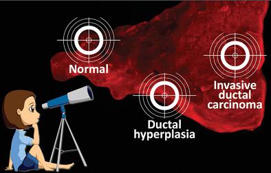
Clean surgical margins in breast-conserving surgery (BCS) are essential for preventing recurrence. Intraoperative pathologic diagnostic methods, such as frozen section analysis and imprint cytology, have been recognized as crucial tools in BCS. However, the complexity and time-consuming nature of these pathologic procedures still inhibit their broader applicability worldwide. To address this situation, two issues should be considered: 1) the development of nonpathologic intraoperative diagnosis methods that have better sensitivity, specificity, speed, and cost; and 2) the promotion of new imaging algorithms to standardize data for analyzing positive margins, as represented by artificial intelligence (AI), without the need for judgment by well-trained pathologists. Researchers have attempted to develop new methods or techniques; several have recently emerged for real-time intraoperative management of breast margins in live tissues. These methods include conventional imaging, spectroscopy, tomography, magnetic resonance imaging, microscopy, fluorescent probes, and multimodal imaging techniques. This work summarizes the traditional pathologic and newly developed techniques and discusses the advantages and disadvantages of each method. Taking into consideration the recent advances in analyzing pathologic data from breast cancer tissue with AI, the combined use of new technologies with AI algorithms is proposed, and future directions for real-time intraoperative margin assessment in BCS are discussed.
中文翻译:

未来保乳手术中实时术中边缘评估的新兴技术。
保乳手术 (BCS) 中清洁的手术边缘对于预防复发至关重要。术中病理诊断方法,如冰冻切片分析和印记细胞学,已被认为是 BCS 的重要工具。然而,这些病理程序的复杂性和耗时性仍然限制了它们在世界范围内更广泛的应用。针对这种情况,需要考虑两个问题:1)开发具有更好敏感性、特异性、速度和成本的非病理性术中诊断方法; 2)推广新的成像算法,以标准化分析阳性切缘的数据,以人工智能(AI)为代表,无需训练有素的病理学家进行判断。研究人员尝试开发新方法或技术;最近出现了一些用于实时术中管理活组织乳房边缘的技术。这些方法包括传统成像、光谱学、断层扫描、磁共振成像、显微镜、荧光探针和多模态成像技术。这项工作总结了传统的病理技术和新开发的技术,并讨论了每种方法的优点和缺点。考虑到利用人工智能分析乳腺癌组织病理数据的最新进展,提出了新技术与人工智能算法的结合使用,并讨论了BCS实时术中切缘评估的未来方向。
更新日期:2020-03-17
中文翻译:
未来保乳手术中实时术中边缘评估的新兴技术。
保乳手术 (BCS) 中清洁的手术边缘对于预防复发至关重要。术中病理诊断方法,如冰冻切片分析和印记细胞学,已被认为是 BCS 的重要工具。然而,这些病理程序的复杂性和耗时性仍然限制了它们在世界范围内更广泛的应用。针对这种情况,需要考虑两个问题:1)开发具有更好敏感性、特异性、速度和成本的非病理性术中诊断方法; 2)推广新的成像算法,以标准化分析阳性切缘的数据,以人工智能(AI)为代表,无需训练有素的病理学家进行判断。研究人员尝试开发新方法或技术;最近出现了一些用于实时术中管理活组织乳房边缘的技术。这些方法包括传统成像、光谱学、断层扫描、磁共振成像、显微镜、荧光探针和多模态成像技术。这项工作总结了传统的病理技术和新开发的技术,并讨论了每种方法的优点和缺点。考虑到利用人工智能分析乳腺癌组织病理数据的最新进展,提出了新技术与人工智能算法的结合使用,并讨论了BCS实时术中切缘评估的未来方向。


 京公网安备 11010802027423号