当前位置:
X-MOL 学术
›
Acta Biomater.
›
论文详情
Our official English website, www.x-mol.net, welcomes your
feedback! (Note: you will need to create a separate account there.)
Biological coating with platelet-rich plasma and adipose tissue-derived microvascular fragments improves the vascularization, biocompatibility and tissue incorporation of porous polyethylene.
Acta Biomaterialia ( IF 9.4 ) Pub Date : 2020-03-17 , DOI: 10.1016/j.actbio.2020.03.018 Thomas Später 1 , Anne L Tobias 1 , Maximilian M Menger 2 , Ruth M Nickels 1 , Michael D Menger 1 , Matthias W Laschke 1
Acta Biomaterialia ( IF 9.4 ) Pub Date : 2020-03-17 , DOI: 10.1016/j.actbio.2020.03.018 Thomas Später 1 , Anne L Tobias 1 , Maximilian M Menger 2 , Ruth M Nickels 1 , Michael D Menger 1 , Matthias W Laschke 1
Affiliation
|
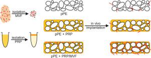
Porous polyethylene (pPE) is a commonly used biomaterial in craniofacial reconstructive surgery. However, implant failure due to insufficient vascularization represents a major issue. To overcome this problem, we herein introduce an effective strategy to improve the vascularization and incorporation of pPE. Adipose tissue-derived microvascular fragments (MVF) from transgenic green fluorescent protein (GFP)+ mice were suspended in platelet-rich plasma (PRP) for the coating of pPE. PRP/MVF-coated pPE as well as PRP-coated and uncoated controls were subsequently implanted into the dorsal skinfold chamber and the flanks of GFP- wild-type mice to analyze their in vivo performance throughout 2, 4 and 8 weeks by means of intravital fluorescence microscopy, histology and immunohistochemistry. The GFP+/GFP- cross-over design allowed the identification of GFP+ MVF within the implants. Shortly after implantation, they rapidly reassembled into new blood-perfused microvascular networks, resulting in a significantly accelerated vascularization of PRP/MVF-coated pPE when compared to both controls. The overall numbers of rolling and adherent leukocytes within the microcirculation as well as macrophages, multi-nucleated giant cells and mast cells around the implants did not differ between the three groups. However, in contrast to uncoated controls, PRP/MVF-coated and PRP-coated pPE promoted pro-angiogenic M2 macrophage polarization at the implantation site. These findings demonstrate that PRP/MVF-coating represents a highly effective strategy to enhance the vascularization, biocompatibility and tissue incorporation of pPE. STATEMENT OF SIGNIFICANCE: The clinical in vivo performance of implanted biomaterials is crucially dependent on their adequate incorporation into the body. To achieve this, we herein introduce an effective biological coating strategy. Our results demonstrate that coating with PRP and MVF accelerates and enhances the vascularization, biocompatibility and tissue incorporation of porous polyethylene. Because this type of biological coating is easily applicable on any type of biomaterial, our approach may rapidly be translated into clinical practice to improve the outcome of various regenerative approaches.
中文翻译:

富含血小板的血浆和脂肪组织衍生的微血管碎片的生物涂层改善了多孔聚乙烯的血管化,生物相容性和组织掺入。
多孔聚乙烯(pPE)是颅面重建手术中常用的生物材料。然而,由于血管化不足引起的植入物失败是一个主要问题。为了克服这个问题,我们在本文中引入了一种有效的策略来改善pPE的血管形成和结合。将来自转基因绿色荧光蛋白(GFP)+小鼠的脂肪组织衍生的微血管片段(MVF)悬浮在富含血小板的血浆(PRP)中,以涂覆pPE。随后将PRP / MVF包被的pPE以及PRP包被的和未包被的对照植入到背部皮褶室和GFP野生型小鼠的腹侧,以通过玻璃体内分析其整个2、4和8周的体内性能荧光显微镜,组织学和免疫组化。GFP + / GFP-交叉设计允许在植入物中鉴定GFP + MVF。植入后不久,它们迅速重新组装成新的血液灌注微血管网络,与两个对照组相比,它们显着加速了PRP / MVF涂层的pPE的血管形成。三组之间,微循环内的滚动白细胞和粘附白细胞的总数以及植入物周围的巨噬细胞,多核巨细胞和肥大细胞没有差异。但是,与未包被的对照相比,PRP / MVF包被和PRP包被的pPE在植入部位促进促血管生成的M2巨噬细胞极化。这些发现表明,PRP / MVF涂层代表了增强pPE的血管形成,生物相容性和组织结合的高效策略。意义声明:植入生物材料的临床体内性能至关重要地取决于它们是否充分掺入人体。为了实现这一点,我们在这里介绍一种有效的生物涂层策略。我们的结果表明,使用PRP和MVF涂层可加速并增强多孔聚乙烯的血管形成,生物相容性和组织掺入。由于这种类型的生物涂层可轻松应用于任何类型的生物材料,因此我们的方法可以迅速转化为临床实践,以改善各种再生方法的效果。我们的结果表明,使用PRP和MVF涂层可加速并增强多孔聚乙烯的血管形成,生物相容性和组织掺入。由于这种类型的生物涂层可轻松应用于任何类型的生物材料,因此我们的方法可以迅速转化为临床实践,以改善各种再生方法的效果。我们的结果表明,用PRP和MVF涂层可加速并增强多孔聚乙烯的血管形成,生物相容性和组织掺入。由于这种类型的生物涂层可轻松应用于任何类型的生物材料,因此我们的方法可以迅速转化为临床实践,以改善各种再生方法的效果。
更新日期:2020-03-17
中文翻译:
富含血小板的血浆和脂肪组织衍生的微血管碎片的生物涂层改善了多孔聚乙烯的血管化,生物相容性和组织掺入。
多孔聚乙烯(pPE)是颅面重建手术中常用的生物材料。然而,由于血管化不足引起的植入物失败是一个主要问题。为了克服这个问题,我们在本文中引入了一种有效的策略来改善pPE的血管形成和结合。将来自转基因绿色荧光蛋白(GFP)+小鼠的脂肪组织衍生的微血管片段(MVF)悬浮在富含血小板的血浆(PRP)中,以涂覆pPE。随后将PRP / MVF包被的pPE以及PRP包被的和未包被的对照植入到背部皮褶室和GFP野生型小鼠的腹侧,以通过玻璃体内分析其整个2、4和8周的体内性能荧光显微镜,组织学和免疫组化。GFP + / GFP-交叉设计允许在植入物中鉴定GFP + MVF。植入后不久,它们迅速重新组装成新的血液灌注微血管网络,与两个对照组相比,它们显着加速了PRP / MVF涂层的pPE的血管形成。三组之间,微循环内的滚动白细胞和粘附白细胞的总数以及植入物周围的巨噬细胞,多核巨细胞和肥大细胞没有差异。但是,与未包被的对照相比,PRP / MVF包被和PRP包被的pPE在植入部位促进促血管生成的M2巨噬细胞极化。这些发现表明,PRP / MVF涂层代表了增强pPE的血管形成,生物相容性和组织结合的高效策略。意义声明:植入生物材料的临床体内性能至关重要地取决于它们是否充分掺入人体。为了实现这一点,我们在这里介绍一种有效的生物涂层策略。我们的结果表明,使用PRP和MVF涂层可加速并增强多孔聚乙烯的血管形成,生物相容性和组织掺入。由于这种类型的生物涂层可轻松应用于任何类型的生物材料,因此我们的方法可以迅速转化为临床实践,以改善各种再生方法的效果。我们的结果表明,使用PRP和MVF涂层可加速并增强多孔聚乙烯的血管形成,生物相容性和组织掺入。由于这种类型的生物涂层可轻松应用于任何类型的生物材料,因此我们的方法可以迅速转化为临床实践,以改善各种再生方法的效果。我们的结果表明,用PRP和MVF涂层可加速并增强多孔聚乙烯的血管形成,生物相容性和组织掺入。由于这种类型的生物涂层可轻松应用于任何类型的生物材料,因此我们的方法可以迅速转化为临床实践,以改善各种再生方法的效果。


 京公网安备 11010802027423号