Our official English website, www.x-mol.net, welcomes your
feedback! (Note: you will need to create a separate account there.)
Evaluation of Direct Grafting Strategies via Trivalent Anchoring for Enabling Lipid Membrane and Cytoskeleton Staining in Expansion Microscopy.
ACS Nano ( IF 15.8 ) Pub Date : 2020-03-16 , DOI: 10.1021/acsnano.9b09259 Gang Wen 1 , Marisa Vanheusden 1 , Aline Acke 1 , Donato Valli 1 , Robert K Neely 2 , Volker Leen 1 , Johan Hofkens 1, 3
ACS Nano ( IF 15.8 ) Pub Date : 2020-03-16 , DOI: 10.1021/acsnano.9b09259 Gang Wen 1 , Marisa Vanheusden 1 , Aline Acke 1 , Donato Valli 1 , Robert K Neely 2 , Volker Leen 1 , Johan Hofkens 1, 3
Affiliation
|
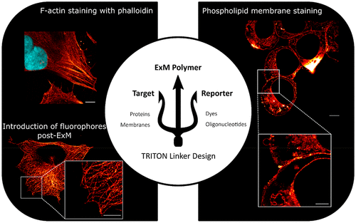
Super-resolution fluorescence microscopy is a key tool in the elucidation of biological fine structures, providing insights into the distribution and interactions of biomolecular complexes down to the nanometer scale. Expansion microscopy is a recently developed approach for achieving nanoscale resolution on a conventional microscope. Here, biological samples are embedded in an isotropically swollen hydrogel. This physical expansion of the sample allows imaging with resolutions down to the tens-of-nanometers. However, because of the requirement that fluorescent labels are covalently bound to the hydrogel, standard, small-molecule targeting of fluorophores has proven incompatible with expansion microscopy. Here, we show a chemical linking approach that enables direct, covalent grafting of a targeting molecule and fluorophore to the hydrogel in expansion microscopy. We show application of this series of molecules in the antibody-free targeting of the cell cytoskeleton and in an example of lipid membrane staining for expansion microscopy. Furthermore, using this trivalent linker strategy, we demonstrate the benefit of introducing fluorescent labels post-expansion by visualizing an immunostaining through fluorescent oligonucleotide hybridization after expanding the polymer. Our probes allow different labeling approaches that are compatible with expansion microscopy.
中文翻译:

在扩展显微镜下,通过三价锚定对脂质膜和细胞骨架染色的直接嫁接策略进行评估。
超分辨率荧光显微镜是阐明生物精细结构的关键工具,可洞察直至纳米级的生物分子复合物的分布和相互作用。扩展显微镜是一种在常规显微镜上实现纳米级分辨率的最新开发方法。在这里,生物样品被包埋在各向同性溶胀的水凝胶中。样品的这种物理膨胀允许以低至几十纳米的分辨率成像。但是,由于要求荧光标记物与水凝胶共价结合,因此荧光团的标准小分子靶向已证明与扩展显微镜不兼容。在这里,我们展示了一种化学链接方法,可实现直接,在扩展显微镜中将靶向分子和荧光团共价接枝到水凝胶上。我们展示了该系列分子在无抗体靶向的细胞骨架中的应用以及在用于扩展显微镜的脂质膜染色的实例中的应用。此外,使用这种三价接头策略,我们展示了在扩增聚合物后通过可视化通过荧光寡核苷酸杂交的免疫染色在扩增后引入荧光标记的好处。我们的探针允许使用与扩展显微镜兼容的不同标记方法。我们通过在扩增聚合物后通过荧光寡核苷酸杂交观察免疫染色来证明在扩增后引入荧光标记的好处。我们的探针允许使用与扩展显微镜兼容的不同标记方法。我们通过在扩增聚合物后通过荧光寡核苷酸杂交观察免疫染色来证明在扩增后引入荧光标记的好处。我们的探针允许使用与扩展显微镜兼容的不同标记方法。
更新日期:2020-03-16
中文翻译:
在扩展显微镜下,通过三价锚定对脂质膜和细胞骨架染色的直接嫁接策略进行评估。
超分辨率荧光显微镜是阐明生物精细结构的关键工具,可洞察直至纳米级的生物分子复合物的分布和相互作用。扩展显微镜是一种在常规显微镜上实现纳米级分辨率的最新开发方法。在这里,生物样品被包埋在各向同性溶胀的水凝胶中。样品的这种物理膨胀允许以低至几十纳米的分辨率成像。但是,由于要求荧光标记物与水凝胶共价结合,因此荧光团的标准小分子靶向已证明与扩展显微镜不兼容。在这里,我们展示了一种化学链接方法,可实现直接,在扩展显微镜中将靶向分子和荧光团共价接枝到水凝胶上。我们展示了该系列分子在无抗体靶向的细胞骨架中的应用以及在用于扩展显微镜的脂质膜染色的实例中的应用。此外,使用这种三价接头策略,我们展示了在扩增聚合物后通过可视化通过荧光寡核苷酸杂交的免疫染色在扩增后引入荧光标记的好处。我们的探针允许使用与扩展显微镜兼容的不同标记方法。我们通过在扩增聚合物后通过荧光寡核苷酸杂交观察免疫染色来证明在扩增后引入荧光标记的好处。我们的探针允许使用与扩展显微镜兼容的不同标记方法。我们通过在扩增聚合物后通过荧光寡核苷酸杂交观察免疫染色来证明在扩增后引入荧光标记的好处。我们的探针允许使用与扩展显微镜兼容的不同标记方法。


 京公网安备 11010802027423号