当前位置:
X-MOL 学术
›
J. Phys. Chem. C
›
论文详情
Our official English website, www.x-mol.net, welcomes your
feedback! (Note: you will need to create a separate account there.)
Formation of SiP2 Nanocrystals Embedded in SiO2 from Phosphorus-Rich SiO1.5 Thin Films
The Journal of Physical Chemistry C ( IF 3.3 ) Pub Date : 2020-03-26 , DOI: 10.1021/acs.jpcc.9b11416 S. Geiskopf 1 , M. Stoffel 1 , X. Devaux 1 , E. André 2 , C. Carteret 2 , A. Bouché 1 , M. Vergnat 1 , H. Rinnert 1
The Journal of Physical Chemistry C ( IF 3.3 ) Pub Date : 2020-03-26 , DOI: 10.1021/acs.jpcc.9b11416 S. Geiskopf 1 , M. Stoffel 1 , X. Devaux 1 , E. André 2 , C. Carteret 2 , A. Bouché 1 , M. Vergnat 1 , H. Rinnert 1
Affiliation
|
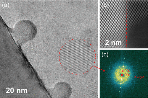
We investigate the structural, vibrational, and optical properties of phosphorus-rich SiO1.5 thin films annealed at 1100 °C. For phosphorus (P) contents larger than 3 atom %, high-resolution transmission electron microscopy characterizations reveal the presence of both spherical-shaped SiP2 nanoparticles crystallizing in an orthorhombic structure and bumps in epitaxy with the underlying Si substrate. Energy-dispersive spectroscopy measurements confirm the SiP2 stoichiometry. Moreover, electron energy loss spectroscopy characterizations allow us to determine the exact location of P and Si atoms. Apart from SiP2 nanoparticles, P atoms are found to be located in the bumps and in the Si substrate to a level of 1 atom %, which is explained by P diffusion during annealing. The vibrational properties determined by Raman spectroscopy are found to be in excellent agreement with density functional theory calculations of the vibration modes for the SiP2 alloy. Finally, the quenching of photoluminescence with an increasing P content is explained on the basis of structural data.
中文翻译:

富含磷的SiO 1.5薄膜在SiO 2中嵌入SiP 2纳米晶体的形成
我们研究了在1100°C退火的富磷SiO 1.5薄膜的结构,振动和光学性质。对于大于3原子%的磷(P)含量,高分辨率透射电子显微镜表征显示,存在以正交晶体结构结晶的球形SiP 2纳米颗粒,以及与下面的Si衬底在外延中凸起的情况。能量色散光谱学测量证实了SiP 2化学计量。此外,电子能量损失光谱学表征使我们能够确定P和Si原子的确切位置。除了SiP 2在纳米粒子中,发现P原子位于凸块和硅衬底中的含量为1原子%,这可以通过退火过程中的P扩散来解释。发现通过拉曼光谱法测定的振动性质与SiP 2合金的振动模式的密度泛函理论计算非常一致。最后,根据结构数据解释了随着P含量增加而猝灭的光致发光。
更新日期:2020-03-27
中文翻译:
富含磷的SiO 1.5薄膜在SiO 2中嵌入SiP 2纳米晶体的形成
我们研究了在1100°C退火的富磷SiO 1.5薄膜的结构,振动和光学性质。对于大于3原子%的磷(P)含量,高分辨率透射电子显微镜表征显示,存在以正交晶体结构结晶的球形SiP 2纳米颗粒,以及与下面的Si衬底在外延中凸起的情况。能量色散光谱学测量证实了SiP 2化学计量。此外,电子能量损失光谱学表征使我们能够确定P和Si原子的确切位置。除了SiP 2在纳米粒子中,发现P原子位于凸块和硅衬底中的含量为1原子%,这可以通过退火过程中的P扩散来解释。发现通过拉曼光谱法测定的振动性质与SiP 2合金的振动模式的密度泛函理论计算非常一致。最后,根据结构数据解释了随着P含量增加而猝灭的光致发光。


 京公网安备 11010802027423号