Our official English website, www.x-mol.net, welcomes your feedback! (Note: you will need to create a separate account there.)
Structural changes in α-chitin through nanofibrillation by high-pressure homogenization in water
Polymer Journal ( IF 2.8 ) Pub Date : 2020-03-13 , DOI: 10.1038/s41428-020-0322-0 Yuko Ono , Kota Ogura , Yuto Kaku , Shuji Fujisawa , Akira Isogai
Polymer Journal ( IF 2.8 ) Pub Date : 2020-03-13 , DOI: 10.1038/s41428-020-0322-0 Yuko Ono , Kota Ogura , Yuto Kaku , Shuji Fujisawa , Akira Isogai
|
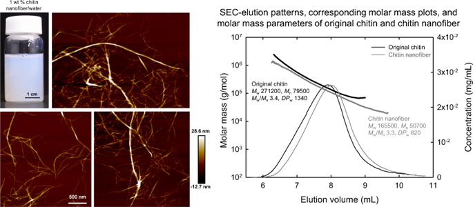
Chitin nanofiber was prepared from purified crab shell chitin by repeated high-pressure homogenization in water. The chitin nanofiber/water dispersion thus prepared was viscous and translucent, and maintained a stable dispersion at room temperature for several months. AFM images showed that the chitin nanofibers had heterogeneous network structures with widths ranging from several nanometers to several tens of nanometers. Some kinks and twisted structures were also observed in the AFM images. X-ray diffraction patterns showed that both the crystallinity index and crystal width of the original alpha-chitin decreased with nanofibrillation. Solid-state 13C-NMR spectra showed that the chemical shifts of all carbons were unchanged before and after nanofibrillation, and that all C6–OH groups had the gauche-gauche conformation irrespective of the crystalline fibril surfaces and insides. The degree of N -acetylation increased from 0.83 to 0.98, which was probably due to C2–NH2 groups present in the original chitin being partially removed during high-pressure homogenization in water. The original chitin and chitin nanofiber were dissolved in 8% LiCl/DMAc, and the solutions after dilution to 1% LiCl/DMAc were subjected to size-exclusion chromatography combined with multi-angle laser-light scattering to determine their molar masses and molar mass distributions. The weight-average molar mass (Mw) value of the original chitin was 271,200 (degree of polymerization [DP] ~1340). The Mw value of the chitin nanofiber was 165,500 (DP 820), showing that the DP of the original chitin decreased by 40% through the nanofibrillation in water to form the chitin nanofiber. Chitin nanofibers were prepared from purified crab-shell chitin particles by repeated high-pressure homogenization in water. AFM images showed that the chitin nanofibers had heterogeneous network structures. X-ray diffraction patterns showed that both the crystallinity index and crystal width of the original α-chitin decreased by nanofibrillation. Solid-state 13 C-NMR spectra showed that all C6–OH groups had the gauche–gauche conformation. The degree of N -acetylation increased from 0.83 to 0.98 by nanofibrillation, while the weight-average molar masses of the original chitin and chitin nanofibers were 271,200 and 165,500, respectively.
中文翻译:

通过在水中高压均质化的纳米纤维化,α-几丁质的结构变化
甲壳素纳米纤维是由纯化的蟹壳甲壳素在水中反复高压均质制备而成的。由此制备的几丁质纳米纤维/水分散体具有粘性和半透明性,并在室温下保持稳定的分散体数月。AFM 图像显示几丁质纳米纤维具有异质网络结构,宽度从几纳米到几十纳米不等。在 AFM 图像中也观察到一些扭结和扭曲结构。X 射线衍射图显示原始 α-几丁质的结晶度指数和晶体宽度都随着纳米纤丝化而降低。固态 13C-NMR 谱表明所有碳的化学位移在纳米纤丝化前后都没有变化,并且所有 C6-OH 基团都具有 gauche-gauche 构象,而与结晶原纤维表面和内部无关。N-乙酰化程度从 0.83 增加到 0.98,这可能是由于在水中高压均质过程中原始几丁质中存在的 C2-NH2 基团被部分去除。将原甲壳素和甲壳素纳米纤维溶解在8% LiCl/DMAc中,稀释至1% LiCl/DMAc后的溶液进行尺寸排阻色谱结合多角度激光散射测定其摩尔质量和摩尔质量分布。原始几丁质的重均摩尔质量 (Mw) 值为 271,200(聚合度 [DP] ~1340)。甲壳素纳米纤维的 Mw 值为 165,500 (DP 820),表明通过在水中纳米纤维化形成几丁质纳米纤维,原始几丁质的 DP 降低了 40%。几丁质纳米纤维是由纯化的蟹壳几丁质颗粒通过在水中反复高压均质制备的。AFM 图像显示几丁质纳米纤维具有异质网络结构。X 射线衍射图显示原始 α-几丁质的结晶度指数和晶体宽度均因纳米纤丝化而降低。固态 13 C-NMR 谱表明所有 C6-OH 基团都具有 gauche-gauche 构象。通过纳米纤维化,N-乙酰化程度从 0.83 增加到 0.98,而原始几丁质和几丁质纳米纤维的重均摩尔质量分别为 271,200 和 165,500。几丁质纳米纤维是由纯化的蟹壳几丁质颗粒通过在水中反复高压均质制备的。AFM 图像显示几丁质纳米纤维具有异质网络结构。X 射线衍射图显示原始 α-几丁质的结晶度指数和晶体宽度均因纳米纤丝化而降低。固态 13 C-NMR 谱表明所有 C6-OH 基团都具有 gauche-gauche 构象。通过纳米纤维化,N-乙酰化程度从 0.83 增加到 0.98,而原始几丁质和几丁质纳米纤维的重均摩尔质量分别为 271,200 和 165,500。几丁质纳米纤维是由纯化的蟹壳几丁质颗粒通过在水中反复高压均质制备的。AFM 图像显示几丁质纳米纤维具有异质网络结构。X 射线衍射图显示原始 α-几丁质的结晶度指数和晶体宽度均因纳米纤丝化而降低。固态 13 C-NMR 谱表明所有 C6-OH 基团都具有 gauche-gauche 构象。通过纳米纤维化,N-乙酰化程度从 0.83 增加到 0.98,而原始几丁质和几丁质纳米纤维的重均摩尔质量分别为 271,200 和 165,500。X 射线衍射图显示原始 α-几丁质的结晶度指数和晶体宽度均因纳米纤丝化而降低。固态 13 C-NMR 谱表明所有 C6-OH 基团都具有 gauche-gauche 构象。通过纳米纤维化,N-乙酰化程度从 0.83 增加到 0.98,而原始几丁质和几丁质纳米纤维的重均摩尔质量分别为 271,200 和 165,500。X 射线衍射图显示原始 α-几丁质的结晶度指数和晶体宽度均因纳米纤丝化而降低。固态 13 C-NMR 谱表明所有 C6-OH 基团都具有 gauche-gauche 构象。通过纳米纤维化,N-乙酰化程度从 0.83 增加到 0.98,而原始几丁质和几丁质纳米纤维的重均摩尔质量分别为 271,200 和 165,500。
更新日期:2020-03-13
中文翻译:
通过在水中高压均质化的纳米纤维化,α-几丁质的结构变化
甲壳素纳米纤维是由纯化的蟹壳甲壳素在水中反复高压均质制备而成的。由此制备的几丁质纳米纤维/水分散体具有粘性和半透明性,并在室温下保持稳定的分散体数月。AFM 图像显示几丁质纳米纤维具有异质网络结构,宽度从几纳米到几十纳米不等。在 AFM 图像中也观察到一些扭结和扭曲结构。X 射线衍射图显示原始 α-几丁质的结晶度指数和晶体宽度都随着纳米纤丝化而降低。固态 13C-NMR 谱表明所有碳的化学位移在纳米纤丝化前后都没有变化,并且所有 C6-OH 基团都具有 gauche-gauche 构象,而与结晶原纤维表面和内部无关。N-乙酰化程度从 0.83 增加到 0.98,这可能是由于在水中高压均质过程中原始几丁质中存在的 C2-NH2 基团被部分去除。将原甲壳素和甲壳素纳米纤维溶解在8% LiCl/DMAc中,稀释至1% LiCl/DMAc后的溶液进行尺寸排阻色谱结合多角度激光散射测定其摩尔质量和摩尔质量分布。原始几丁质的重均摩尔质量 (Mw) 值为 271,200(聚合度 [DP] ~1340)。甲壳素纳米纤维的 Mw 值为 165,500 (DP 820),表明通过在水中纳米纤维化形成几丁质纳米纤维,原始几丁质的 DP 降低了 40%。几丁质纳米纤维是由纯化的蟹壳几丁质颗粒通过在水中反复高压均质制备的。AFM 图像显示几丁质纳米纤维具有异质网络结构。X 射线衍射图显示原始 α-几丁质的结晶度指数和晶体宽度均因纳米纤丝化而降低。固态 13 C-NMR 谱表明所有 C6-OH 基团都具有 gauche-gauche 构象。通过纳米纤维化,N-乙酰化程度从 0.83 增加到 0.98,而原始几丁质和几丁质纳米纤维的重均摩尔质量分别为 271,200 和 165,500。几丁质纳米纤维是由纯化的蟹壳几丁质颗粒通过在水中反复高压均质制备的。AFM 图像显示几丁质纳米纤维具有异质网络结构。X 射线衍射图显示原始 α-几丁质的结晶度指数和晶体宽度均因纳米纤丝化而降低。固态 13 C-NMR 谱表明所有 C6-OH 基团都具有 gauche-gauche 构象。通过纳米纤维化,N-乙酰化程度从 0.83 增加到 0.98,而原始几丁质和几丁质纳米纤维的重均摩尔质量分别为 271,200 和 165,500。几丁质纳米纤维是由纯化的蟹壳几丁质颗粒通过在水中反复高压均质制备的。AFM 图像显示几丁质纳米纤维具有异质网络结构。X 射线衍射图显示原始 α-几丁质的结晶度指数和晶体宽度均因纳米纤丝化而降低。固态 13 C-NMR 谱表明所有 C6-OH 基团都具有 gauche-gauche 构象。通过纳米纤维化,N-乙酰化程度从 0.83 增加到 0.98,而原始几丁质和几丁质纳米纤维的重均摩尔质量分别为 271,200 和 165,500。X 射线衍射图显示原始 α-几丁质的结晶度指数和晶体宽度均因纳米纤丝化而降低。固态 13 C-NMR 谱表明所有 C6-OH 基团都具有 gauche-gauche 构象。通过纳米纤维化,N-乙酰化程度从 0.83 增加到 0.98,而原始几丁质和几丁质纳米纤维的重均摩尔质量分别为 271,200 和 165,500。X 射线衍射图显示原始 α-几丁质的结晶度指数和晶体宽度均因纳米纤丝化而降低。固态 13 C-NMR 谱表明所有 C6-OH 基团都具有 gauche-gauche 构象。通过纳米纤维化,N-乙酰化程度从 0.83 增加到 0.98,而原始几丁质和几丁质纳米纤维的重均摩尔质量分别为 271,200 和 165,500。


 京公网安备 11010802027423号