Redox Biology ( IF 10.7 ) Pub Date : 2020-03-13 , DOI: 10.1016/j.redox.2020.101503 Yongxing Lai 1 , Peiqiang Lin 1 , Manli Chen 2 , Yixian Zhang 3 , Jianhao Chen 2 , Mouwei Zheng 2 , Ji Liu 2 , Houwei Du 2 , Ronghua Chen 2 , Xiaodong Pan 4 , Nan Liu 1 , Hongbin Chen 1
|
Background
Ischemic stroke can induce changes in mitochondrial morphology and function. As a regulatory gene in mitochondria, optic atrophy 1 (OPA1) plays a pivotal role in the regulation of mitochondrial dynamics and other related functions. However, its roles in cerebral ischemia-related conditions are barely understood.
Methods
Cultured rat primary cortical neurons were respectively transfected with OPA1-v1S1-encoding and OPA1-v1-encoding lentivirus before exposure to 2-h oxygen-glucose deprivation (OGD) and subsequent reoxygenation (OGD/R). Adult male SD rats received an intracranial injection of AAV-OPA1-v1S1 and were subjected to 90 min of transient middle cerebral artery occlusion (tMCAO) followed by reperfusion. OPA1 expression and function were detected by in vitro and in vivo assays.
Results
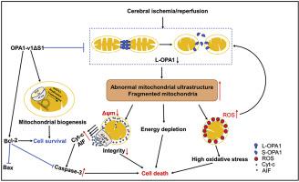
OPA1 was excessively cleaved after cerebral ischemia/reperfusion injury, both in vitro and in vivo. Under OGD/R condition, compared with that of the LV-OPA1-v1-treated group, the expression of OPA1-v1S1 efficiently restored L-OPA1 level and alleviated neuronal death and mitochondrial morphological damage. Meanwhile, the expression of OPA1-v1S1 markedly improved cerebral ischemia/reperfusion-induced motor function damage, attenuated brain infarct volume, neuronal apoptosis, mitochondrial bioenergetics deficits, oxidative stress, and restored the morphology of mitochondrial cristae and mitochondrial length. It also preserved the mitochondrial integrity and reinforced the mtDNA content and expression of mitochondrial biogenesis factors in ischemic rats.
Interpretation
Our results demonstrate that the stabilization of L-OPA1 protects ischemic brains by reducing neuronal apoptosis and preserving mitochondrial function, suggesting its significance as a promising therapeutic target for stroke prevention and treatment.
中文翻译:
撤回:L-OPA1 的恢复通过抑制神经元凋亡和保护线粒体功能减轻大鼠急性缺血性中风损伤
背景
缺血性中风可引起线粒体形态和功能的变化。作为线粒体中的调节基因,视神经萎缩1(OPA1)在线粒体动力学和其他相关功能的调节中发挥着关键作用。然而,它在脑缺血相关疾病中的作用却知之甚少。
方法
培养的大鼠原代皮质神经元分别转染OPA1-v1编码 S1 和 OPA1-v1 的慢病毒在暴露于 2 小时缺氧葡萄糖 (OGD) 和随后的复氧 (OGD/R) 之前。成年雄性 SD 大鼠颅内注射 AAV-OPA1-v1 S1 和接受 90 分钟的短暂大脑中动脉闭塞 (tMCAO),然后再灌注。通过体外和体内测定检测 OPA1 表达和功能。
结果
在脑缺血/再灌注损伤后,OPA1 在体外和体内均被过度裂解。 OGD/R条件下,与LV-OPA1-v1处理组相比,OPA1-v1的表达量S1有效恢复L-OPA1水平并减轻神经元死亡和线粒体形态损伤。同时,OPA1-v1的表达S1显着改善脑缺血/再灌注引起的运动功能损伤,减轻脑梗塞体积,神经元凋亡,线粒体生物能缺陷,氧化应激,并恢复线粒体嵴的形态和线粒体长度。它还保留了缺血大鼠线粒体的完整性并增强了线粒体DNA含量和线粒体生物发生因子的表达。
解释
我们的结果表明,L-OPA1 的稳定通过减少神经元凋亡和保留线粒体功能来保护缺血性大脑,这表明其作为中风预防和治疗的有前景的治疗靶点具有重要意义。


 京公网安备 11010802027423号