当前位置:
X-MOL 学术
›
J. Bone Miner. Res.
›
论文详情
Our official English website, www.x-mol.net, welcomes your
feedback! (Note: you will need to create a separate account there.)
Modeling-Based Bone Formation in the Human Femoral Neck in Subjects Treated With Denosumab.
Journal of Bone and Mineral Research ( IF 5.1 ) Pub Date : 2020-03-12 , DOI: 10.1002/jbmr.4006 David W Dempster 1, 2 , Arkadi Chines 3 , Mathias P Bostrom 4 , Jeri W Nieves 1, 2 , Hua Zhou 2 , Li Chen 3 , Nico Pannacciulli 3 , Rachel B Wagman 3 , Felicia Cosman 1
Journal of Bone and Mineral Research ( IF 5.1 ) Pub Date : 2020-03-12 , DOI: 10.1002/jbmr.4006 David W Dempster 1, 2 , Arkadi Chines 3 , Mathias P Bostrom 4 , Jeri W Nieves 1, 2 , Hua Zhou 2 , Li Chen 3 , Nico Pannacciulli 3 , Rachel B Wagman 3 , Felicia Cosman 1
Affiliation
|
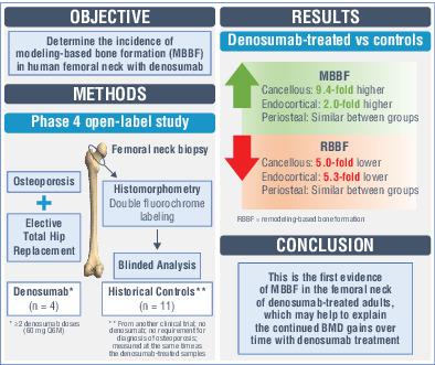
Denosumab is associated with continued gains in hip and spine BMD with up to 10 years of treatment in postmenopausal women with osteoporosis. Despite potent inhibition of bone remodeling, findings in nonhuman primates suggest modeling‐based bone formation (MBBF) may persist during denosumab treatment. This study assessed whether MBBF in the femoral neck (FN) is preserved in the context of inhibited remodeling in subjects receiving denosumab. This open‐label study enrolled postmenopausal women with osteoporosis who had received two or more doses of denosumab (60 mg subcutaneously every 6 months [Q6M]) per standard of care and were planning elective total hip replacement (THR) owing to osteoarthritis of the hip. Transverse sections of the FN were obtained after THR and analyzed histomorphometrically. MBBF, based on fluorochrome labeling and presence of smooth cement lines, was evaluated in cancellous, endocortical, and periosteal envelopes of the FN. Histomorphometric parameters were used to assess MBBF and remodeling‐based bone formation (RBBF) in denosumab‐treated subjects (n = 4; mean age = 73.5 years; range, 70 to 78 years) and historical female controls (n = 11; mean age = 67.8 years; range, 62 to 80 years) obtained from the placebo group of a prior study and not treated with denosumab. All analyses were descriptive. All subjects in both groups exhibited MBBF in the periosteal envelope; in cancellous and endocortical envelopes, all denosumab‐treated subjects and 81.8% of controls showed evidence of MBBF. Compared with controls, denosumab‐treated subjects showed 9.4‐fold and 2.0‐fold higher mean values of MBBF in cancellous and endocortical envelopes, respectively, whereas RBBF mean values were 5.0‐fold and 5.3‐fold lower. In the periosteal envelope, MBBF and RBBF rates were similar between subjects and controls. These results demonstrate the occurrence of MBBF in the human FN and suggest that denosumab preserves MBBF while inhibiting remodeling, which may contribute to the observed continued gains in BMD over time after remodeling is maximally inhibited. © 2020 The Authors. Journal of Bone and Mineral Research published by American Society for Bone and Mineral Research
中文翻译:

接受狄诺塞麦治疗的受试者股骨颈中基于模型的骨形成。
对于患有骨质疏松症的绝经后妇女,经过长达 10 年的治疗,狄诺塞麦与髋部和脊柱 BMD 的持续增加有关。尽管对骨重塑具有有效的抑制作用,但在非人灵长类动物中的研究结果表明,基于模型的骨形成(MBBF)可能在狄诺塞麦治疗期间持续存在。本研究评估了接受狄诺塞麦治疗的受试者在重塑受到抑制的情况下股骨颈 (FN) 中的 MBBF 是否得以保留。这项开放标签研究纳入了患有骨质疏松症的绝经后女性,她们按照标准护理接受了两剂或更多剂量的狄诺塞麦(每 6 个月皮下注射 60 毫克 [Q6M]),并且由于髋部骨关节炎而计划选择性全髋关节置换术 (THR) 。 THR 后获得 FN 的横截面并进行组织形态计量学分析。 MBBF 基于荧光染料标记和光滑水泥线的存在,在 FN 的松质、皮质内和骨膜包膜中进行评估。使用组织形态学参数评估接受狄诺塞麦治疗的受试者( n = 4;平均年龄 = 73.5 岁;范围,70 至 78 岁)和历史女性对照者( n = 11;平均年龄)的 MBBF 和基于重塑的骨形成 (RBBF) = 67.8 岁;范围,62 至 80 岁)取自先前研究的安慰剂组,未接受狄诺塞麦治疗。所有分析都是描述性的。两组中所有受试者的骨膜包膜均表现出 MBBF;在松质和内皮质包膜中,所有接受狄诺塞麦治疗的受试者和 81.8% 的对照者均显示出 MBBF 的证据。与对照组相比,接受狄诺塞麦治疗的受试者松质和皮质内包膜的 MBBF 平均值分别高出 9.4 倍和 2.0 倍,而 RBBF 平均值则低 5.0 倍和 5.3 倍。 在骨膜包膜中,受试者和对照组之间的 MBBF 和 RBBF 率相似。这些结果证明了人 FN 中发生了 MBBF,并表明地诺塞麦在抑制重塑的同时保留了 MBBF,这可能有助于在最大程度地抑制重塑后观察到的 BMD 随着时间的推移持续增加。 © 2020 作者。美国骨与矿物质研究学会出版的《骨与矿物质研究杂志》
更新日期:2020-03-12
中文翻译:
接受狄诺塞麦治疗的受试者股骨颈中基于模型的骨形成。
对于患有骨质疏松症的绝经后妇女,经过长达 10 年的治疗,狄诺塞麦与髋部和脊柱 BMD 的持续增加有关。尽管对骨重塑具有有效的抑制作用,但在非人灵长类动物中的研究结果表明,基于模型的骨形成(MBBF)可能在狄诺塞麦治疗期间持续存在。本研究评估了接受狄诺塞麦治疗的受试者在重塑受到抑制的情况下股骨颈 (FN) 中的 MBBF 是否得以保留。这项开放标签研究纳入了患有骨质疏松症的绝经后女性,她们按照标准护理接受了两剂或更多剂量的狄诺塞麦(每 6 个月皮下注射 60 毫克 [Q6M]),并且由于髋部骨关节炎而计划选择性全髋关节置换术 (THR) 。 THR 后获得 FN 的横截面并进行组织形态计量学分析。 MBBF 基于荧光染料标记和光滑水泥线的存在,在 FN 的松质、皮质内和骨膜包膜中进行评估。使用组织形态学参数评估接受狄诺塞麦治疗的受试者( n = 4;平均年龄 = 73.5 岁;范围,70 至 78 岁)和历史女性对照者( n = 11;平均年龄)的 MBBF 和基于重塑的骨形成 (RBBF) = 67.8 岁;范围,62 至 80 岁)取自先前研究的安慰剂组,未接受狄诺塞麦治疗。所有分析都是描述性的。两组中所有受试者的骨膜包膜均表现出 MBBF;在松质和内皮质包膜中,所有接受狄诺塞麦治疗的受试者和 81.8% 的对照者均显示出 MBBF 的证据。与对照组相比,接受狄诺塞麦治疗的受试者松质和皮质内包膜的 MBBF 平均值分别高出 9.4 倍和 2.0 倍,而 RBBF 平均值则低 5.0 倍和 5.3 倍。 在骨膜包膜中,受试者和对照组之间的 MBBF 和 RBBF 率相似。这些结果证明了人 FN 中发生了 MBBF,并表明地诺塞麦在抑制重塑的同时保留了 MBBF,这可能有助于在最大程度地抑制重塑后观察到的 BMD 随着时间的推移持续增加。 © 2020 作者。美国骨与矿物质研究学会出版的《骨与矿物质研究杂志》


 京公网安备 11010802027423号