PLoS Pathogens ( IF 6.7 ) Pub Date : 2020-03-09 , DOI: 10.1371/journal.ppat.1008392 Jian Shang , Yushun Wan , Chang Liu , Boyd Yount , Kendra Gully , Yang Yang , Ashley Auerbach , Guiqing Peng , Ralph Baric , Fang Li
|
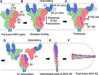
Coronaviruses recognize a variety of receptors using different domains of their envelope-anchored spike protein. How these diverse receptor recognition patterns affect viral entry is unknown. Mouse hepatitis coronavirus (MHV) is the only known coronavirus that uses the N-terminal domain (NTD) of its spike to recognize a protein receptor, CEACAM1a. Here we determined the cryo-EM structure of MHV spike complexed with mouse CEACAM1a. The trimeric spike contains three receptor-binding S1 heads sitting on top of a trimeric membrane-fusion S2 stalk. Three receptor molecules bind to the sides of the spike trimer, where three NTDs are located. Receptor binding induces structural changes in the spike, weakening the interactions between S1 and S2. Using protease sensitivity and negative-stain EM analyses, we further showed that after protease treatment of the spike, receptor binding facilitated the dissociation of S1 from S2, allowing S2 to transition from pre-fusion to post-fusion conformation. Together these results reveal a new role of receptor binding in MHV entry: in addition to its well-characterized role in viral attachment to host cells, receptor binding also induces the conformational change of the spike and hence the fusion of viral and host membranes. Our study provides new mechanistic insight into coronavirus entry and highlights the diverse entry mechanisms used by different viruses.
中文翻译:
小鼠冠状病毒刺突蛋白与受体复合的结构揭示了病毒进入的机制
冠状病毒使用其被膜锚定的刺突蛋白的不同结构域识别多种受体。这些不同的受体识别方式如何影响病毒进入尚不清楚。小鼠肝炎冠状病毒(MHV)是唯一已知的使用冠状病毒N末端结构域(NTD)识别蛋白质受体CEACAM1a的冠状病毒。在这里,我们确定了与小鼠CEACAM1a复合的MHV穗的冷冻EM结构。三聚体刺突包含位于三聚体膜融合S2茎杆顶部的三个结合受体的S1头。三个受体分子结合到三聚体的三聚体的侧面。受体结合诱导尖峰的结构变化,削弱了S1和S2之间的相互作用。使用蛋白酶敏感性和阴性染色EM分析,我们进一步表明,对刺突进行蛋白酶处理后,受体结合促进了S1从S2的解离,使S2从融合前构象转变为融合后构象。这些结果共同揭示了受体结合在MHV进入中的新作用:除了其在病毒附着于宿主细胞中的良好表征的作用外,受体结合还诱导突触的构象变化,从而诱导病毒膜与宿主膜融合。我们的研究提供了有关冠状病毒进入的新机制,并强调了不同病毒使用的各种进入机制。受体结合除了在病毒与宿主细胞的附着中发挥了很好的作用外,还结合了刺突的构象变化,从而导致病毒膜与宿主膜融合。我们的研究提供了有关冠状病毒进入的新机制,并强调了不同病毒使用的各种进入机制。受体结合除了在病毒与宿主细胞的附着中发挥了很好的作用外,还结合了刺突的构象变化,从而导致病毒膜与宿主膜融合。我们的研究提供了有关冠状病毒进入的新机制,并强调了不同病毒使用的各种进入机制。


 京公网安备 11010802027423号