PLoS Pathogens ( IF 5.5 ) Pub Date : 2020-03-09 , DOI: 10.1371/journal.ppat.1008383 Laura Belot 1 , Malika Ouldali 1 , Stéphane Roche 1 , Pierre Legrand 2 , Yves Gaudin 1 , Aurélie A Albertini 1
|
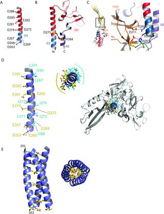
Mokola virus (MOKV) belongs to the lyssavirus genus. As other genus members—including rabies virus (RABV)—it causes deadly encephalitis in mammals. MOKV entry into host cells is mediated by its transmembrane glycoprotein G. First, G binds cellular receptors, triggering virion endocytosis. Then, in the acidic endosomal environment, G undergoes a conformational change from its pre- toward its post-fusion state that catalyzes the merger of the viral and endosomal membranes. Here, we have determined the crystal structure of a soluble MOKV G ectodomain in which the hydrophobic fusion loops have been replaced by more hydrophilic sequences. The crystal structure corresponds to a monomer that is similar to the protomer of the trimeric post-fusion state of vesicular stomatitis virus (VSV) G. However, by electron microscopy, we show that, at low pH, at the surface of pseudotyped VSV, MOKV spikes adopt the trimeric post-fusion conformation and have a tendency to reorganize into regular arrays. Sequence alignment between MOKV G and RABV G allows a precise location of RABV G antigenic sites. Repositioning MOKV G domains on VSV G pre-fusion structure reveals that antigenic sites are located in the most exposed part of the molecule in its pre-fusion conformation and are therefore very accessible to antibodies. Furthermore, the structure allows the identification of pH-sensitive molecular switches. Specifically, the long helix, which constitutes the core of the post-fusion trimer for class III fusion glycoproteins, contains many acidic residues located at the trimeric interface. Several of them, aligned along the helix, point toward the trimer axis. They have to be protonated for the post-fusion trimer to be stable. At high pH, when they are negatively charged, they destabilize the interface, which explains the conformational change reversibility. Finally, the present structure will be of great help to perform rational mutagenesis on lyssavirus glycoproteins.
中文翻译:
Mokola病毒糖蛋白在融合后构象中的晶体结构。
Mokola病毒(MOKV)属于狂犬病病毒属。与狂犬病病毒(RABV)等其他属一样,它会导致哺乳动物致命的脑炎。MOKV进入宿主细胞是由其跨膜糖蛋白G介导的。首先,G结合细胞受体,触发病毒体内吞。然后,在酸性的内体环境中,G经历了从融合前到融合后状态的构象变化,从而催化了病毒膜和内体膜的融合。在这里,我们确定了可溶性MOKV G胞外域的晶体结构,其中疏水融合环已被更多的亲水序列取代。晶体结构对应的单体类似于水泡性口炎病毒(VSV)G的三聚体融合后状态的前体。但是,通过电子显微镜观察,我们发现,在低pH下,在假型VSV的表面,MOKV尖峰采用三聚体融合后构象,并倾向于重组为规则阵列。MOKV G和RABV G之间的序列比对可精确定位RABV G抗原位点。在VSV G融合前结构上重新定位MOKV G结构域显示,抗原位点以融合前构象位于分子最暴露的部分,因此抗体非常容易接近。此外,该结构允许识别pH敏感的分子开关。具体而言,构成III类融合糖蛋白融合后三聚体核心的长螺旋包含位于三聚体界面的许多酸性残基。其中几个沿螺旋线对齐,指向三聚体轴。必须使它们质子化,以使融合后三聚体稳定。在高pH下,当它们带负电荷时,它们会使界面不稳定,这说明了构象变化的可逆性。最后,本结构将对狂犬病病毒糖蛋白进行合理诱变有很大帮助。


 京公网安备 11010802027423号