当前位置:
X-MOL 学术
›
Commun. Biol.
›
论文详情
Our official English website, www.x-mol.net, welcomes your
feedback! (Note: you will need to create a separate account there.)
Spatial and temporal tracking of cardiac exosomes in mouse using a nano-luciferase-CD63 fusion protein.
Communications Biology ( IF 5.2 ) Pub Date : 2020-03-10 , DOI: 10.1038/s42003-020-0830-7 Weijia Luo 1 , Yuan Dai 1 , Zhishi Chen 1 , Xiaojing Yue 2 , Kelsey C Andrade-Powell 1 , Jiang Chang 1
Communications Biology ( IF 5.2 ) Pub Date : 2020-03-10 , DOI: 10.1038/s42003-020-0830-7 Weijia Luo 1 , Yuan Dai 1 , Zhishi Chen 1 , Xiaojing Yue 2 , Kelsey C Andrade-Powell 1 , Jiang Chang 1
Affiliation
|
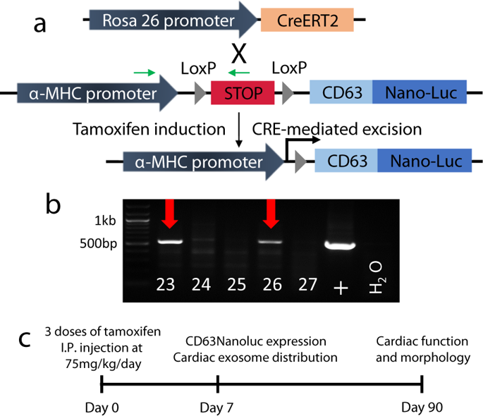
Exosomes are secreted extracellular vesicles with lipid bilayer membranes. They are emerging as a new category of messengers that facilitate cross-talk between cells, tissues, and organs. Thus, a critical demand arises for the development of a sensitive and non-invasive tracking system for endogenous exosomes. We have generated a genetic mouse model that meets this goal. The Nano-luciferase (NanoLuc) reporter was fused with the exosome surface marker CD63 for exosome labeling. The cardiomyocyte-specific αMHC promoter followed by the loxP-STOP-loxP cassette was engineered for temporal and spatial labeling of exosomes originated from cardiomyocytes. The transgenic mouse was bred with a tamoxifen-inducible Cre mouse (Rosa26Cre-ERT2) to achieve inducible expression of CD63NanoLuc reporter. The specific labeling and tissue distribution of endogenous exosomes released from cardiomyocytes were demonstrated by luciferase assay and non-invasive bioluminescent live imaging. This endogenous exosome tracking mouse provides a useful tool for a range of research applications.
中文翻译:

使用纳米荧光素酶-CD63融合蛋白对小鼠心脏外泌体进行时空跟踪。
外泌体是具有脂质双层膜的分泌的细胞外囊泡。它们正在成为使细胞,组织和器官之间发生串扰的一类新型信使。因此,对于开发用于内源性外泌体的灵敏且非侵入性的追踪系统提出了关键需求。我们已经生成了满足此目标的遗传小鼠模型。纳米荧光素酶(NanoLuc)报告基因与外泌体表面标记CD63融合,用于外泌体标记。心肌细胞特异性αMHC启动子,然后是loxP-STOP-loxP盒,经过工程改造,可用于时空标记源自心肌细胞的外泌体。将转基因小鼠与他莫昔芬诱导的Cre小鼠(Rosa26Cre-ERT2)繁殖,以实现CD63NanoLuc报告基因的诱导表达。通过荧光素酶测定和非侵入性生物发光实时成像证实了从心肌细胞释放的内源性外泌体的特异性标记和组织分布。这种内源性外泌体追踪鼠标为一系列研究应用程序提供了有用的工具。
更新日期:2020-03-10
中文翻译:
使用纳米荧光素酶-CD63融合蛋白对小鼠心脏外泌体进行时空跟踪。
外泌体是具有脂质双层膜的分泌的细胞外囊泡。它们正在成为使细胞,组织和器官之间发生串扰的一类新型信使。因此,对于开发用于内源性外泌体的灵敏且非侵入性的追踪系统提出了关键需求。我们已经生成了满足此目标的遗传小鼠模型。纳米荧光素酶(NanoLuc)报告基因与外泌体表面标记CD63融合,用于外泌体标记。心肌细胞特异性αMHC启动子,然后是loxP-STOP-loxP盒,经过工程改造,可用于时空标记源自心肌细胞的外泌体。将转基因小鼠与他莫昔芬诱导的Cre小鼠(Rosa26Cre-ERT2)繁殖,以实现CD63NanoLuc报告基因的诱导表达。通过荧光素酶测定和非侵入性生物发光实时成像证实了从心肌细胞释放的内源性外泌体的特异性标记和组织分布。这种内源性外泌体追踪鼠标为一系列研究应用程序提供了有用的工具。


 京公网安备 11010802027423号