Our official English website, www.x-mol.net, welcomes your
feedback! (Note: you will need to create a separate account there.)
Temporospatial distribution and transcriptional profile of retinal microglia in the oxygen-induced retinopathy mouse model.
Glia ( IF 5.4 ) Pub Date : 2020-03-09 , DOI: 10.1002/glia.23810 Myriam Boeck 1 , Adrian Thien 1 , Julian Wolf 1 , Nora Hagemeyer 2 , Yannik Laich 1 , Dilmurat Yusuf 3 , Rolf Backofen 3 , Peipei Zhang 1 , Stefaniya Boneva 1 , Andreas Stahl 4 , Ingo Hilgendorf 5 , Hansjürgen Agostini 1 , Marco Prinz 2, 6, 7 , Peter Wieghofer 8 , Günther Schlunck 1 , Anja Schlecht 1 , Clemens Lange 1
Glia ( IF 5.4 ) Pub Date : 2020-03-09 , DOI: 10.1002/glia.23810 Myriam Boeck 1 , Adrian Thien 1 , Julian Wolf 1 , Nora Hagemeyer 2 , Yannik Laich 1 , Dilmurat Yusuf 3 , Rolf Backofen 3 , Peipei Zhang 1 , Stefaniya Boneva 1 , Andreas Stahl 4 , Ingo Hilgendorf 5 , Hansjürgen Agostini 1 , Marco Prinz 2, 6, 7 , Peter Wieghofer 8 , Günther Schlunck 1 , Anja Schlecht 1 , Clemens Lange 1
Affiliation
|
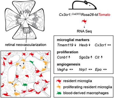
Myeloid cells such as resident retinal microglia (MG) or infiltrating blood‐derived macrophages (Mϕ) accumulate in areas of retinal ischemia and neovascularization (RNV) and modulate neovascular eye disease. Their temporospatial distribution and biological function in this process, however, remain unclarified. Using state‐of‐the‐art methods, including cell‐specific reporter mice and high‐throughput RNA sequencing (RNA Seq), this study determined the extent of MG proliferation and Mϕ infiltration in areas with retinal ischemia and RNV in Cx3cr1 CreERT2:Rosa26‐tdTomato mice and examined the transcriptional profile of MG in the mouse model of oxygen‐induced retinopathy (OIR). For RNA Seq, tdTomato‐positive retinal MG were sorted by flow cytometry followed by Gene ontology (GO) cluster analysis. Furthermore, intraperitoneal injections of the cell proliferation marker 5‐ethynyl‐2′‐deoxyuridine (EdU) were performed from postnatal day (p) 12 to p16. We found that MG is the predominant myeloid cell population while Mϕ rarely appears in areas of RNV. Thirty percent of retinal MG in areas of RNV were EdU‐positive indicating a considerable local MG cell expansion. GO cluster analysis revealed an enrichment of clusters related to cell division, tubulin binding, ATPase activity, protein kinase regulatory activity, and chemokine receptor binding in MG in the OIR model compared to untreated controls. In conclusion, activated retinal MG alter their transcriptional profile, exhibit considerable proliferative ability and are by far the most frequent myeloid cell population in areas of ischemia and RNV in the OIR model thus presenting a potential target for future therapeutic approaches.
中文翻译:

氧诱导视网膜病变小鼠模型中视网膜小胶质细胞的时间空间分布和转录特征。
骨髓细胞,如常驻视网膜小胶质细胞 (MG) 或浸润性血源性巨噬细胞 (Mφ),在视网膜缺血和新生血管 (RNV) 区域积聚,并调节新生血管性眼病。然而,它们在这个过程中的时空分布和生物学功能仍然不清楚。使用最先进的方法,包括细胞特异性报告小鼠和高通量 RNA 测序(RNA Seq),本研究确定了Cx3cr1 CreERT2 中视网膜缺血和 RNV 区域的 MG 增殖和 Mφ 浸润程度:Rosa26 -td番茄小鼠并检查了 MG 在氧诱导视网膜病变 (OIR) 小鼠模型中的转录谱。对于 RNA Seq,tdTomato 阳性视网膜 MG 通过流式细胞术进行排序,然后进行基因本体 (GO) 聚类分析。此外,从出生后第 12 天到第 16 天进行了细胞增殖标记物 5-乙炔基-2'-脱氧尿苷 (EdU) 的腹腔注射。我们发现 MG 是主要的骨髓细胞群,而 Mφ 很少出现在 RNV 区域。RNV 区域 30% 的视网膜 MG 为 EdU 阳性,表明局部 MG 细胞大量扩增。GO 聚类分析显示,与未处理的对照相比,OIR 模型中 MG 中与细胞分裂、微管蛋白结合、ATP 酶活性、蛋白激酶调节活性和趋化因子受体结合相关的簇富集。综上所述,
更新日期:2020-03-09
中文翻译:
氧诱导视网膜病变小鼠模型中视网膜小胶质细胞的时间空间分布和转录特征。
骨髓细胞,如常驻视网膜小胶质细胞 (MG) 或浸润性血源性巨噬细胞 (Mφ),在视网膜缺血和新生血管 (RNV) 区域积聚,并调节新生血管性眼病。然而,它们在这个过程中的时空分布和生物学功能仍然不清楚。使用最先进的方法,包括细胞特异性报告小鼠和高通量 RNA 测序(RNA Seq),本研究确定了Cx3cr1 CreERT2 中视网膜缺血和 RNV 区域的 MG 增殖和 Mφ 浸润程度:Rosa26 -td番茄小鼠并检查了 MG 在氧诱导视网膜病变 (OIR) 小鼠模型中的转录谱。对于 RNA Seq,tdTomato 阳性视网膜 MG 通过流式细胞术进行排序,然后进行基因本体 (GO) 聚类分析。此外,从出生后第 12 天到第 16 天进行了细胞增殖标记物 5-乙炔基-2'-脱氧尿苷 (EdU) 的腹腔注射。我们发现 MG 是主要的骨髓细胞群,而 Mφ 很少出现在 RNV 区域。RNV 区域 30% 的视网膜 MG 为 EdU 阳性,表明局部 MG 细胞大量扩增。GO 聚类分析显示,与未处理的对照相比,OIR 模型中 MG 中与细胞分裂、微管蛋白结合、ATP 酶活性、蛋白激酶调节活性和趋化因子受体结合相关的簇富集。综上所述,


 京公网安备 11010802027423号