Translational Psychiatry ( IF 5.8 ) Pub Date : 2020-03-09 , DOI: 10.1038/s41398-020-0768-z M A Nettis 1, 2 , M Veronese 2, 3 , N Nikkheslat 1 , N Mariani 1 , G Lombardo 1 , L Sforzini 1, 4 , D Enache 5 , N A Harrison 6 , F E Turkheimer 2, 3 , V Mondelli 1, 2 , C M Pariante 1, 2
|
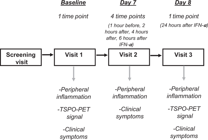
Depression is associated with peripheral inflammation, but its link with brain microglial activity remains unclear. In seven healthy males, we used repeated translocator protein-Positron Emission Tomography (TSPO-PET) dynamic scans with [11C]PBR28 to image brain microglial activation before and 24 h after the immune challenge interferon (IFN)-α. We also investigated the association between changes in peripheral inflammation, changes in microglial activity, and changes in mood. IFN-α administration decreased [11C]PBR28 PET tissue volume of distribution (Vt) across the brain (−20 ± 4%; t6 = 4.1, p = 0.01), but after correction for radioligand free-plasma fraction there were no longer any changes (+23 ± 31%; t = 0.1, p = 0.91). IFN-α increased serum IL-6 (1826 ± 513%, t6 = −7.5, p < 0.001), IL-7 (39 ± 12%, t6 = −3.6, p = 0.01), IL-10 (328 ± 48%, t6 = −12.8, p < 0.001), and IFN-γ (272 ± 64%, t6 = −7.0, p < 0.001) at 4–6 h, and increased serum TNF-α (49 ± 7.6%, t6 = −7.5, p < 0.001), IL-8 (39 ± 12%, t6 = −3.5, p = 0.013), and C-reactive protein (1320 ± 459%, t6 = −7.2, p < 0.001) at 24 h. IFN-α induced temporary mood changes and sickness symptoms after 4–6 h, measured as an increase in POMS-2 total mood score, confusion and fatigue, and a decrease in vigor and friendliness (all p ≤ 0.04). No association was found between changes in peripheral inflammation and changes in PET or mood measures. Our work suggests that brain TSPO-PET signal is highly dependent of inflammation-induced changes in ligand binding to plasma proteins. This limits its usefulness as a sensitive marker of neuroinflammation and consequently, data interpretation. Thus, our results can be interpreted as showing either that [11C]PBR28 is not sensitive enough under these conditions, or that there is simply no microglial activation in this model.
中文翻译:
PET 成像显示健康志愿者接受 IFN-α 免疫攻击后 TSPO 脑密度没有变化。
抑郁症与周围炎症有关,但其与大脑小胶质细胞活动的关系仍不清楚。在七名健康男性中,我们使用[ 11 C]PBR28重复易位蛋白正电子发射断层扫描(TSPO-PET)动态扫描,对免疫攻击干扰素(IFN)-α之前和之后24小时的脑小胶质细胞激活进行成像。我们还研究了外周炎症变化、小胶质细胞活性变化和情绪变化之间的关联。 IFN-α给药降低了[ 11 C]PBR28 PET组织分布体积(Vt)在大脑中的分布(−20 ± 4%;t 6 = 4.1, p = 0.01),但在校正放射性配体游离血浆分数后,没有出现任何变化的时间更长(+23 ± 31%;t = 0.1, p = 0.91)。 IFN-α 增加血清 IL-6 (1826 ± 513%, t 6 = -7.5, p < 0.001)、IL-7 (39 ± 12%, t 6 = -3.6, p = 0.01)、IL-10 ( 328 ± 48%,t 6 = -12.8, p < 0.001),4-6 小时时 IFN-γ (272 ± 64%,t 6 = -7.0, p < 0.001),血清 TNF-α 增加(49 ± 7.6%, t 6 = −7.5, p < 0.001)、IL-8 (39 ± 12%, t 6 = −3.5, p = 0.013) 和 C 反应蛋白 (1320 ± 459%, t 6 = -7.2, p < 0.001) 在 24 小时。 IFN-α 在 4-6 小时后引起暂时的情绪变化和疾病症状,测量为 POMS-2 总情绪评分、困惑和疲劳的增加,以及活力和友善度的下降(所有p ≤ 0.04)。未发现外周炎症的变化与 PET 或情绪测量的变化之间存在关联。 我们的工作表明,大脑 TSPO-PET 信号高度依赖于炎症诱导的配体与血浆蛋白结合的变化。这限制了它作为神经炎症敏感标记的有用性,从而限制了数据解释。因此,我们的结果可以解释为表明[ 11 C]PBR28 在这些条件下不够敏感,或者该模型中根本没有小胶质细胞激活。


 京公网安备 11010802027423号