当前位置:
X-MOL 学术
›
Bioconjugate Chem.
›
论文详情
Our official English website, www.x-mol.net, welcomes your
feedback! (Note: you will need to create a separate account there.)
Live-Cell Imaging at the Nanoscale with Bioconjugatable and Photoactivatable Fluorophores.
Bioconjugate Chemistry ( IF 4.0 ) Pub Date : 2020-03-09 , DOI: 10.1021/acs.bioconjchem.0c00073 Yang Zhang 1 , Françisco M Raymo 1
Bioconjugate Chemistry ( IF 4.0 ) Pub Date : 2020-03-09 , DOI: 10.1021/acs.bioconjchem.0c00073 Yang Zhang 1 , Françisco M Raymo 1
Affiliation
|
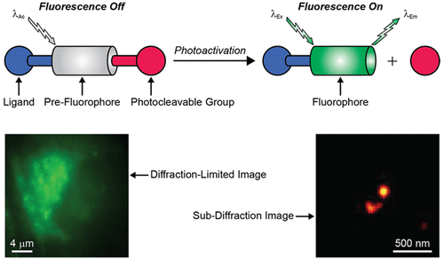
Optical diffraction fundamentally limits the spatial resolution of conventional fluorescence images to length scales that are, at least, 2 orders of magnitude longer than the dimensions of individual molecules. As a result, the development of innovative probes and imaging schemes to overcome diffraction is very much needed to enable the investigation of the fundamental factors regulating cellular functions at the molecular level. In this context, the chemical synthesis of molecular constructs with photoactivatable fluorescence and the ability to label subcellular components of live cells can have transformative implications. Indeed, the fluorescence of the resulting assemblies can be activated with spatiotemporal control, even in the intracellular environment, to permit the sequential localization of individual emissive labels with precision at the nanometer level and the gradual reconstruction of images with subdiffraction resolution. The implementation of these operating principles for subdiffraction imaging, however, is only possible if demanding photochemical and photophysical requirements to enable photoactivation and localization as well as stringent structural requisites to allow the covalent labeling of intracellular targets in live cells are satisfied. Because of these complications, only a few synthetic photoactivatable fluorophores with appropriate performance for live-cell imaging at the nanoscale have been developed so far. Significant synthetic efforts in conjunction with spectroscopic analyses are still very much needed to advance this promising research area further and turn photoactivatable fluorophores into the imaging probes of choice for the investigation of live cells.
中文翻译:

具有可生物缀合和可光活化的荧光团的纳米级活细胞成像。
光学衍射从根本上将常规荧光图像的空间分辨率限制为长度尺度,该尺度至少比单个分子的尺度长2个数量级。结果,迫切需要开发创新的探针和成像方案来克服衍射,以便能够研究在分子水平上调节细胞功能的基本因素。在这种情况下,具有光活化荧光的分子构建体的化学合成以及标记活细胞亚细胞成分的能力可能具有转化意义。实际上,即使在细胞内环境中,也可以通过时空控制来激活所得装配体的荧光,以允许单个发射标记的顺序定位在纳米级别,并逐步重建具有亚衍射分辨率的图像。但是,只有满足苛刻的光化学和光物理要求以实现光激活和定位以及严格的结构要求以允许在活细胞中对细胞内靶标进行共价标记时,才能实现这些用于亚衍射成像的操作原理。由于这些复杂性,到目前为止,仅开发了几种具有适当性能的合成光活化荧光团,用于纳米级的活细胞成像。
更新日期:2020-04-23
中文翻译:
具有可生物缀合和可光活化的荧光团的纳米级活细胞成像。
光学衍射从根本上将常规荧光图像的空间分辨率限制为长度尺度,该尺度至少比单个分子的尺度长2个数量级。结果,迫切需要开发创新的探针和成像方案来克服衍射,以便能够研究在分子水平上调节细胞功能的基本因素。在这种情况下,具有光活化荧光的分子构建体的化学合成以及标记活细胞亚细胞成分的能力可能具有转化意义。实际上,即使在细胞内环境中,也可以通过时空控制来激活所得装配体的荧光,以允许单个发射标记的顺序定位在纳米级别,并逐步重建具有亚衍射分辨率的图像。但是,只有满足苛刻的光化学和光物理要求以实现光激活和定位以及严格的结构要求以允许在活细胞中对细胞内靶标进行共价标记时,才能实现这些用于亚衍射成像的操作原理。由于这些复杂性,到目前为止,仅开发了几种具有适当性能的合成光活化荧光团,用于纳米级的活细胞成像。


 京公网安备 11010802027423号