当前位置:
X-MOL 学术
›
ACS Cent. Sci.
›
论文详情
Our official English website, www.x-mol.net, welcomes your
feedback! (Note: you will need to create a separate account there.)
Live-Cell Imaging and Quantification of PolyQ Aggregates by Stimulated Raman Scattering of Selective Deuterium Labeling.
ACS Central Science ( IF 12.7 ) Pub Date : 2020-03-06 , DOI: 10.1021/acscentsci.9b01196 Kun Miao 1 , Lu Wei 1
ACS Central Science ( IF 12.7 ) Pub Date : 2020-03-06 , DOI: 10.1021/acscentsci.9b01196 Kun Miao 1 , Lu Wei 1
Affiliation
|
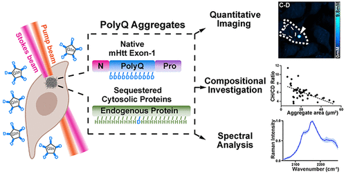
Polyglutamine (polyQ) diseases are a group of neurodegenerative disorders, involving the deposition of aggregation-prone proteins with long polyQ expansions. However, the cytotoxic roles of these aggregates remain highly controversial, largely due to a lack of proper tools for quantitative and nonperturbative interrogations. Common methods including in vitro biochemical, spectroscopic assays, and live-cell fluorescence imaging all suffer from certain limitations. Here, we propose coupling stimulated Raman scattering microscopy with deuterium-labeled glutamine for live-cell imaging, quantification, and spectral analysis of native polyQ aggregates with subcellular resolution. First, through the enrichment of deuterated glutamine in the polyQ sequence of mutant Huntingtin (mHtt) exon1 proteins for Huntington's disease, we achieved sensitive and specific stimulated Raman scattering (SRS) imaging of carbon-deuterium bonds (C-D) from aggregates without GFP labeling, which is commonly employed in fluorescence microscopy. We revealed that these aggregates became 1.8-fold denser compared to those with GFP. Second, we performed ratiometric quantifications, which indicate a surprising dependence of protein compositions on aggregation sizes. Our further calculations, for the first time, reported the absolute concentrations for sequestered mHtt and non-mHtt proteins within the same aggregates. Third, we adopted hyperspectral SRS for Raman spectroscopic studies of aggregate structures. By inducing a cellular heat shock response, a potential therapeutic approach for inhibiting aggregate formation, we found a possible aggregate intermediate state with changed solvation microenvironments. Our method may hence readily unveil new features and mechanistic insight of polyQ aggregates and pave the way for comprehensive in vivo investigations.
中文翻译:

活细胞成像和定量氘标记的刺激拉曼散射对PolyQ聚集体的定量。
聚谷氨酰胺(polyQ)疾病是一组神经退行性疾病,涉及具有长polyQ扩展的易于聚集蛋白的沉积。但是,这些聚集体的细胞毒性作用仍然存在争议,主要是由于缺乏用于定量和非干扰性询问的适当工具。包括体外生化,光谱测定法和活细胞荧光成像在内的常用方法均存在某些局限性。在这里,我们提出耦合刺激的拉曼散射显微镜与氘标记的谷氨酰胺的活细胞成像,量化和光谱分析天然polyQ聚集体与亚细胞分辨率。首先,通过在突变的Huntingtin(mHtt)exon1蛋白的polyQ序列中富集氘化的谷氨酰胺来治疗Huntington病,我们获得了未使用GFP标记的聚集体中碳-氘键(CD)的灵敏和特异激发拉曼散射(SRS)成像,该成像通常在荧光显微镜下使用。我们发现与GFP相比,这些聚集体的密度提高了1.8倍。其次,我们进行了比例定量,这表明蛋白质组成对聚集大小的出乎意料的依赖性。我们的进一步计算首次报告了同一聚集物中隔离的mHtt和非mHtt蛋白的绝对浓度。第三,我们采用高光谱SRS进行聚集体结构的拉曼光谱研究。通过诱导细胞热休克反应,一种抑制聚集体形成的潜在治疗方法,我们发现了可能的聚集中间状态,其溶剂化微环境发生了变化。因此,我们的方法可能会轻易揭示polyQ聚集体的新功能和机理见解,并为全面的体内研究铺平道路。
更新日期:2020-04-23
中文翻译:
活细胞成像和定量氘标记的刺激拉曼散射对PolyQ聚集体的定量。
聚谷氨酰胺(polyQ)疾病是一组神经退行性疾病,涉及具有长polyQ扩展的易于聚集蛋白的沉积。但是,这些聚集体的细胞毒性作用仍然存在争议,主要是由于缺乏用于定量和非干扰性询问的适当工具。包括体外生化,光谱测定法和活细胞荧光成像在内的常用方法均存在某些局限性。在这里,我们提出耦合刺激的拉曼散射显微镜与氘标记的谷氨酰胺的活细胞成像,量化和光谱分析天然polyQ聚集体与亚细胞分辨率。首先,通过在突变的Huntingtin(mHtt)exon1蛋白的polyQ序列中富集氘化的谷氨酰胺来治疗Huntington病,我们获得了未使用GFP标记的聚集体中碳-氘键(CD)的灵敏和特异激发拉曼散射(SRS)成像,该成像通常在荧光显微镜下使用。我们发现与GFP相比,这些聚集体的密度提高了1.8倍。其次,我们进行了比例定量,这表明蛋白质组成对聚集大小的出乎意料的依赖性。我们的进一步计算首次报告了同一聚集物中隔离的mHtt和非mHtt蛋白的绝对浓度。第三,我们采用高光谱SRS进行聚集体结构的拉曼光谱研究。通过诱导细胞热休克反应,一种抑制聚集体形成的潜在治疗方法,我们发现了可能的聚集中间状态,其溶剂化微环境发生了变化。因此,我们的方法可能会轻易揭示polyQ聚集体的新功能和机理见解,并为全面的体内研究铺平道路。


 京公网安备 11010802027423号