JAMA Dermatology ( IF 11.5 ) Pub Date : 2020-05-01 , DOI: 10.1001/jamadermatol.2020.0059 Ashlee M Margheim 1 , Emily C McKenzie 2
|
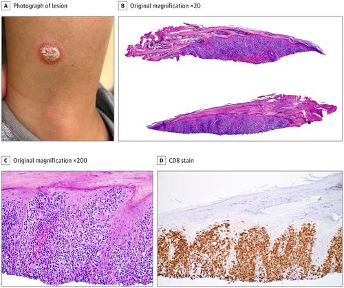
A preschool-age girl presented for evaluation of an asymptomatic plaque on the right lower extremity that had been present for 1 year. Prior treatments included triamcinolone ointment and frankincense with no improvement. A potassium hydroxide preparation had negative results for fungal elements. Her medical history and family history were unremarkable. An examination found a 1-cm nummular plaque with centralized micaceous scale and a smooth erythematous border on the right lateral lower leg (Figure, A). A tangential-shave biopsy was performed to characterize the lesion. Histopathological testing revealed an epidermis featuring elongated rete ridges, keratinocyte enlargement, and a few Civatte bodies, with overlying parakeratotic crust and neutrophilic cell debris. Marked epidermotropism of atypical lymphocytes with irregular nuclear contours and halos, both singly and in small collections (Pautrier microabscesses) were also observed (Figure, B and C). Also, lymphocytes were present along the epidermal side of the dermoepidermal junction. Periodic acid–Schiff stained sections failed to show fungal or yeast elements. Immunohistochemical staining was markedly positive for CD3, CD5, and CD8 (Figure, D); CD4 reactivity was less robust and mainly restricted to dermal lymphocytes. Also, CD20 was present in 5% of dermal lymphocytes, and CD30 showed variable staining of 5% of lymphocytes.
中文翻译:
一个年轻女孩下肢的孤立鳞状斑块。
一名学龄前女孩出现,以评估已经存在1年的右下肢无症状斑块。先前的治疗包括曲安西龙软膏和乳香,但无改善。氢氧化钾制剂对真菌元素的检测结果为阴性。她的病史和家族病史均不明显。检查发现1厘米的环形斑块具有集中的云母鳞片,右小腿外侧有光滑的红斑边界(图A)。进行切向剃刮活检以表征病变。组织病理学检查显示表皮具有细长的网状脊,角质形成细胞增大和一些Civatte体,上面有角化不全的结皮和嗜中性细胞碎片。还观察到非典型淋巴细胞的明显表皮趋化性,其单个和少量集合(Pautrier微脓肿)均具有不规则的核轮廓和光晕(图B和C)。另外,沿真皮表皮连接处的表皮侧存在淋巴细胞。高碘酸-席夫(Schiff)染色切片未显示真菌或酵母元素。免疫组织化学染色对CD3,CD5和CD8呈阳性(图D)。CD4反应性较弱,主要限于真皮淋巴细胞。同样,CD20存在于5%的真皮淋巴细胞中,而CD30显示出5%的淋巴细胞可变染色。高碘酸-席夫(Schiff)染色切片未显示真菌或酵母元素。免疫组化染色显示CD3,CD5和CD8呈阳性(图D);CD4反应性较弱,主要限于真皮淋巴细胞。同样,CD20存在于5%的真皮淋巴细胞中,而CD30显示出5%的淋巴细胞可变染色。高碘酸-席夫(Schiff)染色切片未显示真菌或酵母元素。免疫组化染色显示CD3,CD5和CD8呈阳性(图D);CD4反应性较弱,主要限于真皮淋巴细胞。同样,CD20存在于5%的真皮淋巴细胞中,而CD30显示出5%的淋巴细胞可变染色。


 京公网安备 11010802027423号