当前位置:
X-MOL 学术
›
J. Struct. Biol.
›
论文详情
Our official English website, www.x-mol.net, welcomes your
feedback! (Note: you will need to create a separate account there.)
Automated cryo-lamella preparation for high-throughput in-situ structural biology.
Journal of Structural Biology ( IF 3.0 ) Pub Date : 2020-02-29 , DOI: 10.1016/j.jsb.2020.107488 Genevieve Buckley 1 , Gediminas Gervinskas 2 , Cyntia Taveneau 1 , Hariprasad Venugopal 2 , James C Whisstock 3 , Alex de Marco 4
Journal of Structural Biology ( IF 3.0 ) Pub Date : 2020-02-29 , DOI: 10.1016/j.jsb.2020.107488 Genevieve Buckley 1 , Gediminas Gervinskas 2 , Cyntia Taveneau 1 , Hariprasad Venugopal 2 , James C Whisstock 3 , Alex de Marco 4
Affiliation
|
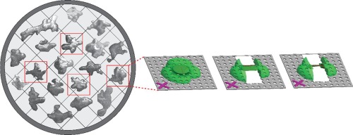
Cryo-transmission electron tomography (cryo-ET) in association with cryo-focused ion beam (cryo-FIB) milling enables structural biology studies to be performed directly within the cellular environment. Cryo-preserved cells are milled and a lamella with a typical thickness of 200-300 nm provides an electron transparent window suitable for cryo-ET imaging. Cryo-FIB milling is an effective method, but it is a tedious and time-consuming process, which typically results in ~10 lamellae per day. Here, we introduce an automated method to reproducibly prepare cryo-lamellae on a grid and reduce the amount of human supervision. We tested the routine on cryo-preserved Saccharomyces cerevisiae, mammalian 293 T cells, and lysozyme protein crystals. Here we demonstrate that our method allows an increased throughput, achieving a rate of 5 lamellae/hour without the need to supervise the FIB milling. We demonstrate that the quality of the lamellae is consistent throughout the preparation and their compatibility with cryo-ET analyses.
中文翻译:

用于高通量原位结构生物学的自动冷冻薄片制备。
低温透射电子层析成像(cryo-ET)与低温聚焦离子束(cryo-FIB)研磨相结合,使结构生物学研究可以直接在细胞环境中进行。将冷冻保存的细胞磨碎,典型厚度为200-300 nm的薄层可提供适用于cryo-ET成像的电子透明窗口。低温FIB研磨是一种有效的方法,但它是一个繁琐且耗时的过程,通常每天会产生约10个薄片。在这里,我们介绍了一种自动化方法,可重复地在网格上制备冷冻薄片,并减少了人工监督的工作量。我们在冷冻保存的酿酒酵母,哺乳动物293 T细胞和溶菌酶蛋白晶体上测试了常规程序。在这里,我们证明了我们的方法可以提高吞吐量,无需监督FIB铣削即可达到每小时5片的速度。我们证明了薄片的质量在整个制备过程中是一致的,并且与cryo-ET分析兼容。
更新日期:2020-03-26
中文翻译:
用于高通量原位结构生物学的自动冷冻薄片制备。
低温透射电子层析成像(cryo-ET)与低温聚焦离子束(cryo-FIB)研磨相结合,使结构生物学研究可以直接在细胞环境中进行。将冷冻保存的细胞磨碎,典型厚度为200-300 nm的薄层可提供适用于cryo-ET成像的电子透明窗口。低温FIB研磨是一种有效的方法,但它是一个繁琐且耗时的过程,通常每天会产生约10个薄片。在这里,我们介绍了一种自动化方法,可重复地在网格上制备冷冻薄片,并减少了人工监督的工作量。我们在冷冻保存的酿酒酵母,哺乳动物293 T细胞和溶菌酶蛋白晶体上测试了常规程序。在这里,我们证明了我们的方法可以提高吞吐量,无需监督FIB铣削即可达到每小时5片的速度。我们证明了薄片的质量在整个制备过程中是一致的,并且与cryo-ET分析兼容。


 京公网安备 11010802027423号