当前位置:
X-MOL 学术
›
Int. J. Numer. Method. Biomed. Eng.
›
论文详情
Our official English website, www.x-mol.net, welcomes your
feedback! (Note: you will need to create a separate account there.)
Automated segmentation of abdominal organs from contrast-enhanced computed tomography using analysis of texture features.
International Journal for Numerical Methods in Biomedical Engineering ( IF 2.2 ) Pub Date : 2020-03-03 , DOI: 10.1002/cnm.3309 Alexander Danilov 1, 2, 3 , Alexandra Yurova 1, 2
International Journal for Numerical Methods in Biomedical Engineering ( IF 2.2 ) Pub Date : 2020-03-03 , DOI: 10.1002/cnm.3309 Alexander Danilov 1, 2, 3 , Alexandra Yurova 1, 2
Affiliation
|
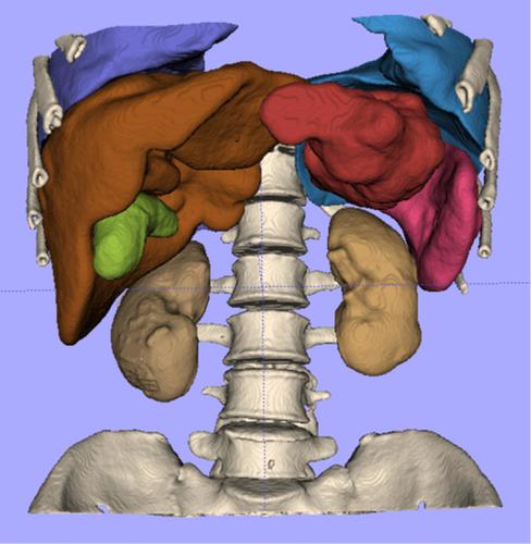
Generation of three‐dimensional personalized geometric models of anatomical structures is an important process for many practical tasks: computer‐aided diagnosis, treatment planning and numerical modeling in biomedical applications. Despite many efforts done by different research groups, automatic segmentation of organs still does not have any general solution. The main difficulties are caused by peculiarities of different medical imaging modalities, image variability (for the same modality) resulting from the wide range of imaging devices, noise and artifacts, large patient anatomical variability and overlapping of intensity ranges of neighboring anatomical structures. In this article, we propose segmentation method based on analysis of texture features and developed specially for segmentation of abdominal organs. Its main advantage is robustness to interpatient gray level and anatomical variability. The proposed method was validated on the patient data. The method implementation was accelerated using graphics processing unit (GPU).
中文翻译:

使用纹理特征分析从对比增强的计算机断层扫描中自动分割腹部器官。
三维解剖结构的个性化几何模型的生成是许多实际任务的重要过程:计算机辅助诊断,治疗计划和生物医学应用中的数值建模。尽管不同研究小组做出了许多努力,但器官的自动分割仍然没有任何通用的解决方案。主要困难是由于不同的医学成像方式的特殊性,由于成像设备范围广,噪声和伪影,大的患者解剖变异性以及相邻解剖结构的强度范围重叠而导致的图像变异性(对于同一形式)。在本文中,我们提出了一种基于纹理特征分析的分割方法,专门针对腹部器官的分割而开发。它的主要优点是对患者间灰度水平和解剖结构可变性的鲁棒性。对患者数据验证了所提出的方法。使用图形处理单元(GPU)加速了该方法的实现。
更新日期:2020-03-03
中文翻译:
使用纹理特征分析从对比增强的计算机断层扫描中自动分割腹部器官。
三维解剖结构的个性化几何模型的生成是许多实际任务的重要过程:计算机辅助诊断,治疗计划和生物医学应用中的数值建模。尽管不同研究小组做出了许多努力,但器官的自动分割仍然没有任何通用的解决方案。主要困难是由于不同的医学成像方式的特殊性,由于成像设备范围广,噪声和伪影,大的患者解剖变异性以及相邻解剖结构的强度范围重叠而导致的图像变异性(对于同一形式)。在本文中,我们提出了一种基于纹理特征分析的分割方法,专门针对腹部器官的分割而开发。它的主要优点是对患者间灰度水平和解剖结构可变性的鲁棒性。对患者数据验证了所提出的方法。使用图形处理单元(GPU)加速了该方法的实现。


 京公网安备 11010802027423号