当前位置:
X-MOL 学术
›
J. Struct. Biol.
›
论文详情
Our official English website, www.x-mol.net, welcomes your
feedback! (Note: you will need to create a separate account there.)
Direct comparison of optical and electron microscopy methods for structural characterization of extracellular vesicles.
Journal of Structural Biology ( IF 3.0 ) Pub Date : 2020-02-04 , DOI: 10.1016/j.jsb.2020.107474 Jade M Noble 1 , LaDeidra Monét Roberts 2 , Netta Vidavsky 3 , Aaron E Chiou 2 , Claudia Fischbach 4 , Matthew J Paszek 5 , Lara A Estroff 6 , Lena F Kourkoutis 7
Journal of Structural Biology ( IF 3.0 ) Pub Date : 2020-02-04 , DOI: 10.1016/j.jsb.2020.107474 Jade M Noble 1 , LaDeidra Monét Roberts 2 , Netta Vidavsky 3 , Aaron E Chiou 2 , Claudia Fischbach 4 , Matthew J Paszek 5 , Lara A Estroff 6 , Lena F Kourkoutis 7
Affiliation
|
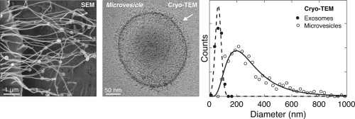
As interest in the role of extracellular vesicles in cell-to-cell communication has increased, so has the use of microscopy and analytical techniques to assess their formation, release, and morphology. In this study, we evaluate scanning electron microscopy (SEM) and cryo-SEM for characterizing the formation and shedding of vesicles from human breast cell lines, parental and hyaluronan synthase 3-(HAS3)-overexpressing MCF10A cells, grown directly on transmission electron microscopy (TEM) grids. While cells imaged with conventional and cryo-SEM exhibit distinct morphologies due to the sample preparation process for each technique, tubular structures protruding from the cell surfaces were observed with both approaches. For HAS3-MCF10A cells, vesicles were present along the length of membrane protrusions. Once completely shed from the cells, extracellular vesicles were characterized using nanoparticle tracking analysis (NTA) and cryo-TEM. The size distributions obtained by each technique were different not only in the range of vesicles analyzed, but also in the relative proportion of smaller-to-larger vesicles. These differences are attributed to the presence of biological debris in the media, which is difficult to differentiate from vesicles in NTA. Furthermore, we demonstrate that cryo-TEM can be used to distinguish between vesicles based on their respective surface structures, thereby providing a path to differentiating vesicle subpopulations and identifying their size distributions. Our study emphasizes the necessity of pairing several techniques to characterize extracellular vesicles.
中文翻译:

直接比较光学和电子显微镜方法对细胞外囊泡结构表征的影响。
随着人们对细胞外囊泡在细胞间通讯中的作用的兴趣不断增加,使用显微镜和分析技术来评估其形成、释放和形态也随之增加。在本研究中,我们评估扫描电子显微镜 (SEM) 和冷冻 SEM 来表征直接在透射电子显微镜上生长的人乳腺细胞系、亲本和透明质酸合酶 3-(HAS3) 过表达 MCF10A 细胞的囊泡形成和脱落(TEM)网格。虽然由于每种技术的样品制备过程,使用传统扫描电镜和冷冻扫描电镜成像的细胞表现出不同的形态,但两种方法都观察到从细胞表面突出的管状结构。对于 HAS3-MCF10A 细胞,沿着膜突起的长度存在囊泡。一旦细胞外囊泡完全从细胞中脱落,就可以使用纳米颗粒跟踪分析 (NTA) 和冷冻透射电镜对细胞外囊泡进行表征。每种技术获得的尺寸分布不仅在分析的囊泡范围上不同,而且在较小与较大囊泡的相对比例上也不同。这些差异归因于培养基中存在生物碎片,很难将其与 NTA 中的囊泡区分开来。此外,我们证明冷冻-TEM 可用于根据囊泡各自的表面结构来区分囊泡,从而提供区分囊泡亚群并确定其大小分布的途径。我们的研究强调了将几种技术配对来表征细胞外囊泡的必要性。
更新日期:2020-03-26
中文翻译:
直接比较光学和电子显微镜方法对细胞外囊泡结构表征的影响。
随着人们对细胞外囊泡在细胞间通讯中的作用的兴趣不断增加,使用显微镜和分析技术来评估其形成、释放和形态也随之增加。在本研究中,我们评估扫描电子显微镜 (SEM) 和冷冻 SEM 来表征直接在透射电子显微镜上生长的人乳腺细胞系、亲本和透明质酸合酶 3-(HAS3) 过表达 MCF10A 细胞的囊泡形成和脱落(TEM)网格。虽然由于每种技术的样品制备过程,使用传统扫描电镜和冷冻扫描电镜成像的细胞表现出不同的形态,但两种方法都观察到从细胞表面突出的管状结构。对于 HAS3-MCF10A 细胞,沿着膜突起的长度存在囊泡。一旦细胞外囊泡完全从细胞中脱落,就可以使用纳米颗粒跟踪分析 (NTA) 和冷冻透射电镜对细胞外囊泡进行表征。每种技术获得的尺寸分布不仅在分析的囊泡范围上不同,而且在较小与较大囊泡的相对比例上也不同。这些差异归因于培养基中存在生物碎片,很难将其与 NTA 中的囊泡区分开来。此外,我们证明冷冻-TEM 可用于根据囊泡各自的表面结构来区分囊泡,从而提供区分囊泡亚群并确定其大小分布的途径。我们的研究强调了将几种技术配对来表征细胞外囊泡的必要性。


 京公网安备 11010802027423号