当前位置:
X-MOL 学术
›
Stem Cells Transl. Med.
›
论文详情
Our official English website, www.x-mol.net, welcomes your
feedback! (Note: you will need to create a separate account there.)
Exosomal 2',3'-CNP from mesenchymal stem cells promotes hippocampus CA1 neurogenesis/neuritogenesis and contributes to rescue of cognition/learning deficiencies of damaged brain.
STEM CELLS Translational Medicine ( IF 5.4 ) Pub Date : 2020-01-15 , DOI: 10.1002/sctm.19-0174 Shih-Yin Chen,Meng-Chieh Lin,Jia-Shiuan Tsai,Pei-Lin He,Wen-Ting Luo,Ing-Ming Chiu,Harvey R Herschman,Hua-Jung Li
STEM CELLS Translational Medicine ( IF 5.4 ) Pub Date : 2020-01-15 , DOI: 10.1002/sctm.19-0174 Shih-Yin Chen,Meng-Chieh Lin,Jia-Shiuan Tsai,Pei-Lin He,Wen-Ting Luo,Ing-Ming Chiu,Harvey R Herschman,Hua-Jung Li
|
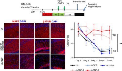
Mesenchymal stem cells (MSCs) have been used in clinical studies to treat neurological diseases and damage. However, implanted MSCs do not achieve their regenerative effects by differentiating into and replacing neural cells. Instead, MSC secretome components mediate the regenerative effects of MSCs. MSC‐derived extracellular vesicles (EVs)/exosomes carry cargo responsible for rescuing brain damage. We previously showed that EP4 antagonist‐induced MSC EVs/exosomes have enhanced regenerative potential to rescue hippocampal damage, compared with EVs/exosomes from untreated MSCs. Here we show that EP4 antagonist‐induced MSC EVs/exosomes promote neurosphere formation in vitro and increase neurogenesis and neuritogenesis in damaged hippocampi; basal MSC EVs/exosomes do not contribute to these regenerative effects. 2′,3′‐Cyclic nucleotide 3′‐phosphodiesterase (CNP) levels in EP4 antagonist‐induced MSC EVs/exosomes are 20‐fold higher than CNP levels in basal MSC EVs/exosomes. Decreasing elevated exosomal CNP levels in EP4 antagonist‐induced MSC EVs/exosomes reduced the efficacy of these EVs/exosomes in promoting β3‐tubulin polymerization and in converting toxic 2′,3′‐cAMP into neuroprotective adenosine. CNP‐depleted EP4 antagonist‐induced MSC EVs/exosomes lost the ability to promote neurogenesis and neuritogenesis in damaged hippocampi. Systemic administration of EV/exosomes from EP4‐antagonist derived MSC EVs/exosomes repaired cognition, learning, and memory deficiencies in mice caused by hippocampal damage. In contrast, CNP‐depleted EP4 antagonist‐induced MSC EVs/exosomes failed to repair this damage. Exosomal CNP contributes to the ability of EP4 antagonist‐elicited MSC EVs/exosomes to promote neurogenesis and neuritogenesis in damaged hippocampi and recovery of cognition, memory, and learning. This experimental approach should be generally applicable to identifying the role of EV/exosomal components in eliciting a variety of biological responses.
中文翻译:

来自间充质干细胞的外泌体2',3'-CNP促进海马CA1神经发生/神经发生,并有助于挽救受损脑的认知/学习缺陷。
间充质干细胞(MSCs)已用于临床研究中,以治疗神经系统疾病和损伤。但是,植入的MSC不能通过分化为神经细胞并替代神经细胞来实现其再生作用。取而代之的是,MSC分泌组的成分介导了MSC的再生作用。MSC衍生的细胞外囊泡(EVs / exosome)携带负责挽救脑损伤的货物。我们以前表明,与未经处理的MSC的EV /外泌体相比,EP 4拮抗剂诱导的MSC EV /外泌体具有增强的修复海马损伤的再生潜力。我们在这里展示了EP 4拮抗剂诱导的MSC EV /外泌体在体外促进神经球形成,并增加受损海马体的神经发生和神经发生。基础MSC EV /外泌体对这些再生作用无贡献。EP 4拮抗剂诱导的MSC EV /外泌体中2',3'-环核苷酸3'-磷酸二酯酶(CNP)的水平比基础MSC EV /外泌体中的CNP高20倍。在EP 4拮抗剂诱导的MSC EV /外泌体中降低外泌体CNP水平升高会降低这些EV /外泌体在促进β3-微管蛋白聚合以及将有毒2',3'-cAMP转化为神经保护腺苷的功效。CNP耗尽的EP 4拮抗剂诱导的MSC EV /外泌体丧失了在受损海马中促进神经发生和神经发生的能力。从EP 4拮抗剂衍生的MSC EV /外泌体系统施用EV /外泌体可修复小鼠海马体损伤引起的认知,学习和记忆缺陷。相反,CNP耗尽的EP 4拮抗剂诱导的MSC EV /外泌体未能修复这种损害。外泌体CNP有助于EP 4拮抗剂诱发的MSC EV /外泌体在受损海马中促进神经发生和神经发生的能力,并恢复认知,记忆和学习能力。这种实验方法通常应适用于确定EV /外泌体成分在引发各种生物学反应中的作用。
更新日期:2020-01-15
中文翻译:
来自间充质干细胞的外泌体2',3'-CNP促进海马CA1神经发生/神经发生,并有助于挽救受损脑的认知/学习缺陷。
间充质干细胞(MSCs)已用于临床研究中,以治疗神经系统疾病和损伤。但是,植入的MSC不能通过分化为神经细胞并替代神经细胞来实现其再生作用。取而代之的是,MSC分泌组的成分介导了MSC的再生作用。MSC衍生的细胞外囊泡(EVs / exosome)携带负责挽救脑损伤的货物。我们以前表明,与未经处理的MSC的EV /外泌体相比,EP 4拮抗剂诱导的MSC EV /外泌体具有增强的修复海马损伤的再生潜力。我们在这里展示了EP 4拮抗剂诱导的MSC EV /外泌体在体外促进神经球形成,并增加受损海马体的神经发生和神经发生。基础MSC EV /外泌体对这些再生作用无贡献。EP 4拮抗剂诱导的MSC EV /外泌体中2',3'-环核苷酸3'-磷酸二酯酶(CNP)的水平比基础MSC EV /外泌体中的CNP高20倍。在EP 4拮抗剂诱导的MSC EV /外泌体中降低外泌体CNP水平升高会降低这些EV /外泌体在促进β3-微管蛋白聚合以及将有毒2',3'-cAMP转化为神经保护腺苷的功效。CNP耗尽的EP 4拮抗剂诱导的MSC EV /外泌体丧失了在受损海马中促进神经发生和神经发生的能力。从EP 4拮抗剂衍生的MSC EV /外泌体系统施用EV /外泌体可修复小鼠海马体损伤引起的认知,学习和记忆缺陷。相反,CNP耗尽的EP 4拮抗剂诱导的MSC EV /外泌体未能修复这种损害。外泌体CNP有助于EP 4拮抗剂诱发的MSC EV /外泌体在受损海马中促进神经发生和神经发生的能力,并恢复认知,记忆和学习能力。这种实验方法通常应适用于确定EV /外泌体成分在引发各种生物学反应中的作用。


 京公网安备 11010802027423号