当前位置:
X-MOL 学术
›
Microsc. Res. Tech.
›
论文详情
Our official English website, www.x-mol.net, welcomes your
feedback! (Note: you will need to create a separate account there.)
A study of digital image analysis on the acrylamide derivative monomers induced apoptosis in rat cerebrum.
Microscopy Research and Technique ( IF 2.0 ) Pub Date : 2020-01-08 , DOI: 10.1002/jemt.23431 Serpil Ünver Saraydin 1 , Dursun Saraydin 2 , Zeynep Deniz Şahin İnan 1
Microscopy Research and Technique ( IF 2.0 ) Pub Date : 2020-01-08 , DOI: 10.1002/jemt.23431 Serpil Ünver Saraydin 1 , Dursun Saraydin 2 , Zeynep Deniz Şahin İnan 1
Affiliation
|
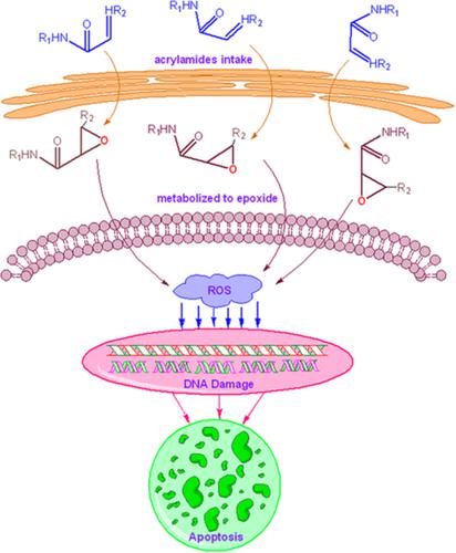
Nowadays, apoptosis is mostly evaluated visually in histological studies. By using the quantitative digital image analysis, this study aimed to investigate the effect of acrylamide‐based monomers (acrylamide [AAm], methacrylamide [MAAm], N‐isopropylacrylamide [NIPAm]) on the cerebrum tissues in rats, which are the most common water‐soluble monomers in the production of polymeric hydrogels used as biomaterials. The Wistar albino rats weighing ~220–240 g were divided into control and three test groups. The control group received 1 mL of saline, and the test groups received 1 mL of aqueous 50 mg/kg/day intramuscular injection of AAm, MAAm, and NIPAm, respectively. At the end of the experiments, brain tissues of all rats euthanized by intramuscular injection of sodium pentobarbital were removed. Terminal deoxynucleotide transferase dUTP nick and labeling (TUNEL) method was applied to brain tissue sections. The monomers have been shown to cause apoptosis due to oxidative stress in cerebrum tissue. Based on apoptosis by tunneling method, quantitative digital image analysis of cell fragments was performed with Olympus cellSens Dimension 1.15 software, and the number, total count area, selected area, average area, and ROI% values of the fragments were found. In addition, the total area and ROI% values of the fragments increased linearly with increasing the molar mass of monomers from the digital image analysis data. Quantitative digital image analysis can facilitate the monitoring of apoptosis caused by the oxidative stress of monomers used in the production of the biomaterials.
中文翻译:

丙烯酰胺衍生物单体诱导大鼠大脑细胞凋亡的数字图像分析研究。
如今,凋亡在组织学研究中大多是通过视觉评估的。通过使用定量数字图像分析,本研究旨在研究最常见的丙烯酰胺基单体(丙烯酰胺[AAm],甲基丙烯酰胺[MAAm],N-异丙基丙烯酰胺[NIPAm])对大鼠大脑组织的影响。用作生物材料的聚合物水凝胶生产中的水溶性单体。体重约220–240 g的Wistar白化病大鼠分为对照组和三个测试组。对照组接受1 mL盐水,测试组分别接受1 mL 50 mg / kg /天的肌内注射Aam,MAAm和NIPAm。在实验结束时,通过肌肉注射戊巴比妥钠安乐死的所有大鼠的脑组织被去除。末端脱氧核苷酸转移酶dUTP切口和标记(TUNEL)方法应用于脑组织切片。已经显示出该单体由于在大脑组织中的氧化应激而引起细胞凋亡。基于隧穿方法的细胞凋亡,使用Olympus cellSens Dimension 1.15软件对细胞片段进行定量数字图像分析,并找到片段的数量,总计数面积,选定面积,平均面积和ROI%值。另外,随着数字图像分析数据中单体摩尔质量的增加,片段的总面积和ROI%值线性增加。定量数字图像分析可以促进对由生物材料生产中使用的单体的氧化应激引起的细胞凋亡进行监测。已经显示出该单体由于在大脑组织中的氧化应激而引起细胞凋亡。基于隧穿方法的细胞凋亡,使用Olympus cellSens Dimension 1.15软件对细胞片段进行定量数字图像分析,并找到片段的数量,总计数面积,选定面积,平均面积和ROI%值。另外,随着数字图像分析数据中单体摩尔质量的增加,片段的总面积和ROI%值线性增加。定量数字图像分析可以促进对由生物材料生产中使用的单体的氧化应激引起的细胞凋亡进行监测。已经表明,由于大脑组织中的氧化应激,单体会引起细胞凋亡。基于隧穿方法的细胞凋亡,使用Olympus cellSens Dimension 1.15软件对细胞片段进行定量数字图像分析,并找到片段的数量,总计数面积,选定面积,平均面积和ROI%值。另外,随着数字图像分析数据中单体摩尔质量的增加,片段的总面积和ROI%值线性增加。定量数字图像分析可以促进对由生物材料生产中使用的单体的氧化应激引起的细胞凋亡进行监测。使用Olympus cellSens Dimension 1.15软件对细胞碎片进行定量数字图像分析,然后发现碎片的数量,总计数面积,选定面积,平均面积和ROI%值。另外,随着数字图像分析数据中单体摩尔质量的增加,片段的总面积和ROI%值线性增加。定量数字图像分析可以促进对由生物材料生产中使用的单体的氧化应激引起的细胞凋亡进行监测。使用Olympus cellSens Dimension 1.15软件对细胞碎片进行定量数字图像分析,然后发现碎片的数量,总计数面积,选定面积,平均面积和ROI%值。另外,随着数字图像分析数据中单体摩尔质量的增加,片段的总面积和ROI%值线性增加。定量数字图像分析可以促进对由生物材料生产中使用的单体的氧化应激引起的细胞凋亡进行监测。片段的总面积和ROI%值随数字图像分析数据中单体的摩尔质量的增加而线性增加。定量数字图像分析可以促进对由生物材料生产中使用的单体的氧化应激引起的细胞凋亡进行监测。片段的总面积和ROI%值随数字图像分析数据中单体的摩尔质量的增加而线性增加。定量数字图像分析可以促进对由生物材料生产中使用的单体的氧化应激引起的细胞凋亡进行监测。
更新日期:2020-01-08
中文翻译:
丙烯酰胺衍生物单体诱导大鼠大脑细胞凋亡的数字图像分析研究。
如今,凋亡在组织学研究中大多是通过视觉评估的。通过使用定量数字图像分析,本研究旨在研究最常见的丙烯酰胺基单体(丙烯酰胺[AAm],甲基丙烯酰胺[MAAm],N-异丙基丙烯酰胺[NIPAm])对大鼠大脑组织的影响。用作生物材料的聚合物水凝胶生产中的水溶性单体。体重约220–240 g的Wistar白化病大鼠分为对照组和三个测试组。对照组接受1 mL盐水,测试组分别接受1 mL 50 mg / kg /天的肌内注射Aam,MAAm和NIPAm。在实验结束时,通过肌肉注射戊巴比妥钠安乐死的所有大鼠的脑组织被去除。末端脱氧核苷酸转移酶dUTP切口和标记(TUNEL)方法应用于脑组织切片。已经显示出该单体由于在大脑组织中的氧化应激而引起细胞凋亡。基于隧穿方法的细胞凋亡,使用Olympus cellSens Dimension 1.15软件对细胞片段进行定量数字图像分析,并找到片段的数量,总计数面积,选定面积,平均面积和ROI%值。另外,随着数字图像分析数据中单体摩尔质量的增加,片段的总面积和ROI%值线性增加。定量数字图像分析可以促进对由生物材料生产中使用的单体的氧化应激引起的细胞凋亡进行监测。已经显示出该单体由于在大脑组织中的氧化应激而引起细胞凋亡。基于隧穿方法的细胞凋亡,使用Olympus cellSens Dimension 1.15软件对细胞片段进行定量数字图像分析,并找到片段的数量,总计数面积,选定面积,平均面积和ROI%值。另外,随着数字图像分析数据中单体摩尔质量的增加,片段的总面积和ROI%值线性增加。定量数字图像分析可以促进对由生物材料生产中使用的单体的氧化应激引起的细胞凋亡进行监测。已经表明,由于大脑组织中的氧化应激,单体会引起细胞凋亡。基于隧穿方法的细胞凋亡,使用Olympus cellSens Dimension 1.15软件对细胞片段进行定量数字图像分析,并找到片段的数量,总计数面积,选定面积,平均面积和ROI%值。另外,随着数字图像分析数据中单体摩尔质量的增加,片段的总面积和ROI%值线性增加。定量数字图像分析可以促进对由生物材料生产中使用的单体的氧化应激引起的细胞凋亡进行监测。使用Olympus cellSens Dimension 1.15软件对细胞碎片进行定量数字图像分析,然后发现碎片的数量,总计数面积,选定面积,平均面积和ROI%值。另外,随着数字图像分析数据中单体摩尔质量的增加,片段的总面积和ROI%值线性增加。定量数字图像分析可以促进对由生物材料生产中使用的单体的氧化应激引起的细胞凋亡进行监测。使用Olympus cellSens Dimension 1.15软件对细胞碎片进行定量数字图像分析,然后发现碎片的数量,总计数面积,选定面积,平均面积和ROI%值。另外,随着数字图像分析数据中单体摩尔质量的增加,片段的总面积和ROI%值线性增加。定量数字图像分析可以促进对由生物材料生产中使用的单体的氧化应激引起的细胞凋亡进行监测。片段的总面积和ROI%值随数字图像分析数据中单体的摩尔质量的增加而线性增加。定量数字图像分析可以促进对由生物材料生产中使用的单体的氧化应激引起的细胞凋亡进行监测。片段的总面积和ROI%值随数字图像分析数据中单体的摩尔质量的增加而线性增加。定量数字图像分析可以促进对由生物材料生产中使用的单体的氧化应激引起的细胞凋亡进行监测。


 京公网安备 11010802027423号