当前位置:
X-MOL 学术
›
STEM CELLS
›
论文详情
Our official English website, www.x-mol.net, welcomes your feedback! (Note: you will need to create a separate account there.)
Pathway-specific reporter genes to study stem cell biology
STEM CELLS ( IF 5.2 ) Pub Date : 2020-03-28 , DOI: 10.1002/stem.3167 Karen M Peterson 1 , Federico Franchi 1 , Michaela Olthoff 1 , Ian Y Chen 2, 3 , Ramasamy Paulmurugan 3, 4 , Martin Rodriguez-Porcel 1
STEM CELLS ( IF 5.2 ) Pub Date : 2020-03-28 , DOI: 10.1002/stem.3167 Karen M Peterson 1 , Federico Franchi 1 , Michaela Olthoff 1 , Ian Y Chen 2, 3 , Ramasamy Paulmurugan 3, 4 , Martin Rodriguez-Porcel 1
Affiliation
|
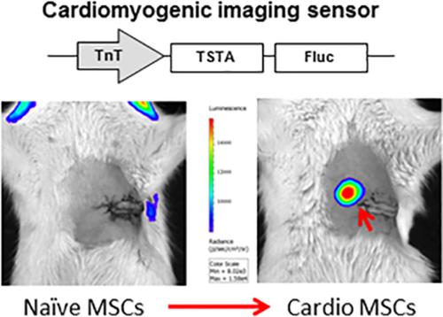
Little is known on the phenotypic characteristics of stem cells (SCs) after they are transplanted to the myocardium, in part due to lack of noninvasive platforms to study SCs directly in the living subject. Reporter gene imaging has played a valuable role in the noninvasive assessment of cell fate in vivo. In this study, we validated a pathway‐specific reporter gene that can be used to noninvasively image the phenotype of SCs transplanted to the myocardium. Rat mesenchymal SCs (MSCs) were studied for phenotypic evidence of myogenic characteristics under in vitro conditions. After markers of myogenic characteristics were identified, we constructed a reporter gene sensor, comprising the firefly luciferase (Fluc) reporter gene driven by the troponin T (TnT) promoter (cardio MSCs had threefold expression in polymerase chain reaction compared to control MSCs) using a two‐step signal amplification strategy. MSCs transfected with TnT‐Fluc were studied and validated under in vitro conditions, showing a strong signal after MSCs acquired myogenic characteristics. Lastly, we observed that cardio MSCs had higher expression of the reporter sensor compared to control cells (0.005 ± 0.0005 vs 0.0025 ± 0.0008 Tnt‐Fluc/ubiquitin‐Fluc, P < .05), and that this novel sensor can detect the change in the phenotype of MSCs directly in the living subject. Pathway‐specific reporter gene imaging allows assessment of changes in the phenotype of MSCs after delivery to the ischemic myocardium, providing important information on the phenotype of these cells. Imaging sensors like the one described here are critical to better understanding of the changes that SCs undergo after transplantation.
中文翻译:

用于研究干细胞生物学的通路特异性报告基因
干细胞 (SCs) 移植到心肌后的表型特征知之甚少,部分原因是缺乏直接在活体受试者中研究 SCs 的非侵入性平台。报告基因成像在体内细胞命运的无创评估中发挥了重要作用。在这项研究中,我们验证了一种通路特异性报告基因,该基因可用于对移植到心肌的 SCs 的表型进行非侵入性成像。研究了大鼠间充质干细胞 (MSC) 在体外条件下的肌原性特征的表型证据。确定肌原性特征标记后,我们构建了一个报告基因传感器,包含由肌钙蛋白 T (TnT) 启动子驱动的萤火虫荧光素酶 (Fluc) 报告基因(与对照 MSC 相比,心脏 MSC 在聚合酶链反应中具有三倍表达),使用两步信号放大策略。在体外条件下对转染 TnT-Fluc 的 MSC 进行了研究和验证,在 MSC 获得肌原性特征后显示出强信号。最后,我们观察到心脏 MSC 与对照细胞相比具有更高的报告传感器表达(0.005 ± 0.0005 vs 0.0025 ± 0.0008 Tnt-Fluc/ubiquitin-Fluc,P < .05),并且这种新型传感器可以检测到直接在活体受试者中的 MSC 表型。通路特异性报告基因成像允许评估输送到缺血心肌后 MSCs 表型的变化,提供有关这些细胞表型的重要信息。像这里描述的那样的成像传感器对于更好地了解 SCs 在移植后经历的变化至关重要。
更新日期:2020-03-28
中文翻译:
用于研究干细胞生物学的通路特异性报告基因
干细胞 (SCs) 移植到心肌后的表型特征知之甚少,部分原因是缺乏直接在活体受试者中研究 SCs 的非侵入性平台。报告基因成像在体内细胞命运的无创评估中发挥了重要作用。在这项研究中,我们验证了一种通路特异性报告基因,该基因可用于对移植到心肌的 SCs 的表型进行非侵入性成像。研究了大鼠间充质干细胞 (MSC) 在体外条件下的肌原性特征的表型证据。确定肌原性特征标记后,我们构建了一个报告基因传感器,包含由肌钙蛋白 T (TnT) 启动子驱动的萤火虫荧光素酶 (Fluc) 报告基因(与对照 MSC 相比,心脏 MSC 在聚合酶链反应中具有三倍表达),使用两步信号放大策略。在体外条件下对转染 TnT-Fluc 的 MSC 进行了研究和验证,在 MSC 获得肌原性特征后显示出强信号。最后,我们观察到心脏 MSC 与对照细胞相比具有更高的报告传感器表达(0.005 ± 0.0005 vs 0.0025 ± 0.0008 Tnt-Fluc/ubiquitin-Fluc,P < .05),并且这种新型传感器可以检测到直接在活体受试者中的 MSC 表型。通路特异性报告基因成像允许评估输送到缺血心肌后 MSCs 表型的变化,提供有关这些细胞表型的重要信息。像这里描述的那样的成像传感器对于更好地了解 SCs 在移植后经历的变化至关重要。


 京公网安备 11010802027423号