Kidney International ( IF 14.8 ) Pub Date : 2020-03-03 , DOI: 10.1016/j.kint.2020.02.011 Qiyang Chen 1 , Jaesok Yu 1 , Brittney M Rush 2 , Sean D Stocker 2 , Roderick J Tan 2 , Kang Kim 3
|
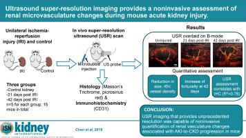
Acute kidney injury (AKI) is a risk factor for the development of chronic kidney disease (CKD). One mechanism for this phenomenon is renal microvascular rarefaction and subsequent chronic impairment in perfusion. However, diagnostic tools to monitor the renal microvasculature in a noninvasive and quantitative manner are still lacking. Ultrasound super-resolution imaging is an emerging technology that can identify microvessels with unprecedented resolution. Here, we applied this imaging technique to identify microvessels in the unilateral ischemia-reperfusion injury mouse model of AKI-to-CKD progression in vivo. Kidneys from 21 and 42 day post- ischemia-reperfusion injury, the contralateral uninjured kidneys, and kidneys from sham-operated mice were examined by ultrasound super-resolution and histology. Renal microvessels were successfully identified by this imaging modality with a resolution down to 32 μm. Renal fibrosis was observed in all kidneys with ischemia-reperfusion injury and was associated with a significant reduction in kidney size, cortical thickness, relative blood volume, and microvascular density as assessed by this imaging. Tortuosity of the cortical microvasculature was also significantly increased at 42 days compared to sham. These vessel density measurements correlated significantly with CD31 immunohistochemistry (R2=0.77). Thus, ultrasound super-resolution imaging provides unprecedented resolution and is capable of noninvasive quantification of renal vasculature changes associated with AKI-to-CKD progression in mice. Hence, this technique could be a promising diagnostic tool for monitoring progressive kidney disease.
中文翻译:
超声超分辨率成像提供了小鼠急性肾损伤期间肾脏微血管变化的无创评估。
急性肾损伤 (AKI) 是慢性肾病 (CKD) 发展的危险因素。这种现象的一种机制是肾微血管稀疏和随后的灌注慢性损伤。然而,仍然缺乏以非侵入性和定量方式监测肾脏微血管的诊断工具。超声超分辨率成像是一项新兴技术,能够以前所未有的分辨率识别微血管。在这里,我们应用这种成像技术来识别体内AKI 到 CKD 进展的单侧缺血再灌注损伤小鼠模型中的微血管. 通过超声超分辨率和组织学检查来自缺血再灌注损伤后第 21 天和第 42 天的肾脏、对侧未受伤肾脏和假手术小鼠的肾脏。通过这种成像方式成功识别了肾脏微血管,分辨率低至 32 μm。在所有缺血再灌注损伤的肾脏中都观察到了肾纤维化,并且与通过该成像评估的肾脏大小、皮质厚度、相对血容量和微血管密度的显着减小有关。与假手术相比,42 天时皮质微血管的曲折度也显着增加。这些血管密度测量值与 CD31 免疫组织化学显着相关 (R 2=0.77)。因此,超声超分辨率成像提供了前所未有的分辨率,并且能够无创量化与小鼠 AKI 到 CKD 进展相关的肾血管变化。因此,该技术可能是监测进行性肾病的一种很有前途的诊断工具。


 京公网安备 11010802027423号