当前位置:
X-MOL 学术
›
Eur. J. Pharm. Sci.
›
论文详情
Our official English website, www.x-mol.net, welcomes your feedback! (Note: you will need to create a separate account there.)
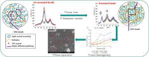
Evolution of the microstructure and the drug release upon annealing the drug loaded lipid-surfactant microspheres.
European Journal of Pharmaceutical Sciences ( IF 4.6 ) Pub Date : 2020-03-02 , DOI: 10.1016/j.ejps.2020.105278 Varun Kushwah 1 , Diogo Gomes Lopes 1 , Ioannis Koutsamanis 1 , Harald Plank 2 , Ioan Ardelean 3 , Avik Sarkar 4 , Andrew Prpich 4 , Mary T Am Ende 4 , Heather Frericks Schmidt 4 , Pankaj Doshi 4 , Sheri L Shamblin 4 , Peter Laggner 1 , Amrit Paudel 5
European Journal of Pharmaceutical Sciences ( IF 4.6 ) Pub Date : 2020-03-02 , DOI: 10.1016/j.ejps.2020.105278 Varun Kushwah 1 , Diogo Gomes Lopes 1 , Ioannis Koutsamanis 1 , Harald Plank 2 , Ioan Ardelean 3 , Avik Sarkar 4 , Andrew Prpich 4 , Mary T Am Ende 4 , Heather Frericks Schmidt 4 , Pankaj Doshi 4 , Sheri L Shamblin 4 , Peter Laggner 1 , Amrit Paudel 5
Affiliation
|
The present study investigates the drug release-governing microstructural properties of melt spray congealed microspheres encapsulating the drug crystals in the matrix of glyceryl behenate and poloxamer (pore former). The solid-state, morphology, and micromeritics of the microspheres were characterized, before and after annealing, using calorimetry, X-ray scattering, porosimetry, scanning electron microscopy, and, NMR diffusometry. The in vitro drug release from and water uptake by the microspheres were obtained. The extent and the rate of drug release from the microspheres increased with a high poloxamer content and at higher annealing temperature and RH. All the drug release profiles were describable using the Higuchi release kinetics pointing towards the diffusion controlled release, both before and after annealing. The annealing process led to the polymorphic conversion of lipid and the increase in the pore size, predominantly at a higher temperature and humidity and for a high poloxamer content. The poloxamer domain increased from an initial 300 nm, up to 2000 nm upon annealing. The water diffusion rate inside the annealed microsphere was twice as fast as for unannealed counterparts. The findings relate the overall phase and pore structure change of the microsphere to the increased drug release induced by annealing. This work serves as a basis for the rational understanding of the modification of the in vitro performance by annealing, a widely used post-process for solid lipid products.
中文翻译:

在将载有药物的脂质表面活性剂微球退火后,微观结构的演变和药物释放。
本研究研究了将药物晶体包封在山hen酸甘油酯和泊洛沙姆(孔形成剂)的基质中的熔融喷雾凝固微球的控制药物释放的微结构特性。在退火之前和之后,使用量热法,X射线散射,孔隙率法,扫描电子显微镜和NMR扩散法对微球的固态,形态和微商进行了表征。获得了微球的体外药物释放和水吸收。随着泊洛沙姆含量高和在较高的退火温度和相对湿度下,药物从微球中释放的程度和速率增加。在退火之前和之后,均使用指向扩散控制释放的Higuchi释放动力学来描述所有药物的释放曲线。退火过程导致脂质的多晶型转化和孔尺寸的增加,主要是在较高的温度和湿度下以及较高的泊洛沙姆含量下。泊洛沙姆域从最初的300 nm增加到退火时的2000 nm。退火微球内部的水扩散速度是未退火对应物的两倍。这些发现将微球的整体相和孔结构变化与退火引起的药物释放增加有关。这项工作为合理理解通过退火(广泛用于固体脂质产品的后处理)对体外性能的改变提供了基础。泊洛沙姆域从最初的300 nm增加到退火时的2000 nm。退火微球内部的水扩散速度是未退火对应物的两倍。这些发现将微球的整体相和孔结构变化与退火引起的药物释放增加有关。这项工作为合理理解通过退火(广泛用于固体脂质产品的后处理)对体外性能的改变提供了基础。泊洛沙姆域从最初的300 nm增加到退火时的2000 nm。退火微球内部的水扩散速度是未退火对应物的两倍。这些发现将微球的整体相和孔结构变化与退火引起的药物释放增加有关。这项工作为合理理解通过退火(广泛用于固体脂质产品的后处理)对体外性能的改变提供了基础。
更新日期:2020-03-03
中文翻译:
在将载有药物的脂质表面活性剂微球退火后,微观结构的演变和药物释放。
本研究研究了将药物晶体包封在山hen酸甘油酯和泊洛沙姆(孔形成剂)的基质中的熔融喷雾凝固微球的控制药物释放的微结构特性。在退火之前和之后,使用量热法,X射线散射,孔隙率法,扫描电子显微镜和NMR扩散法对微球的固态,形态和微商进行了表征。获得了微球的体外药物释放和水吸收。随着泊洛沙姆含量高和在较高的退火温度和相对湿度下,药物从微球中释放的程度和速率增加。在退火之前和之后,均使用指向扩散控制释放的Higuchi释放动力学来描述所有药物的释放曲线。退火过程导致脂质的多晶型转化和孔尺寸的增加,主要是在较高的温度和湿度下以及较高的泊洛沙姆含量下。泊洛沙姆域从最初的300 nm增加到退火时的2000 nm。退火微球内部的水扩散速度是未退火对应物的两倍。这些发现将微球的整体相和孔结构变化与退火引起的药物释放增加有关。这项工作为合理理解通过退火(广泛用于固体脂质产品的后处理)对体外性能的改变提供了基础。泊洛沙姆域从最初的300 nm增加到退火时的2000 nm。退火微球内部的水扩散速度是未退火对应物的两倍。这些发现将微球的整体相和孔结构变化与退火引起的药物释放增加有关。这项工作为合理理解通过退火(广泛用于固体脂质产品的后处理)对体外性能的改变提供了基础。泊洛沙姆域从最初的300 nm增加到退火时的2000 nm。退火微球内部的水扩散速度是未退火对应物的两倍。这些发现将微球的整体相和孔结构变化与退火引起的药物释放增加有关。这项工作为合理理解通过退火(广泛用于固体脂质产品的后处理)对体外性能的改变提供了基础。


 京公网安备 11010802027423号