当前位置:
X-MOL 学术
›
J. Biophotonics
›
论文详情
Our official English website, www.x-mol.net, welcomes your
feedback! (Note: you will need to create a separate account there.)
Optimal imaging windows of indocyanine green-assisted near-infrared dental imaging with rat model and its comparison to X-ray imaging.
Journal of Biophotonics ( IF 2.0 ) Pub Date : 2020-03-09 , DOI: 10.1002/jbio.201960232 Zhongqiang Li 1 , Thomas Hartzler 1 , Alexandra Ramos 2 , Michelle L Osborn 2 , Yanping Li 3 , Shaomian Yao 2 , Jian Xu 1
Journal of Biophotonics ( IF 2.0 ) Pub Date : 2020-03-09 , DOI: 10.1002/jbio.201960232 Zhongqiang Li 1 , Thomas Hartzler 1 , Alexandra Ramos 2 , Michelle L Osborn 2 , Yanping Li 3 , Shaomian Yao 2 , Jian Xu 1
Affiliation
|
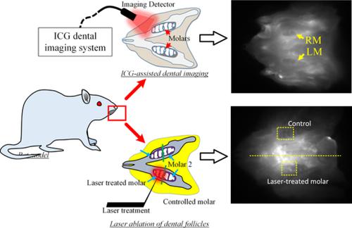
In this study, we used rat animal model to compare the efficiency of indocyanine green (ICG)‐assisted dental near‐infrared fluorescence imaging with X‐ray imaging, and we optimized the imaging window for both unerupted and erupted molars. The results show that the morphology of the dental structures was observed clearly from ICG‐assisted dental images (especially through the endoscope). A better image contrast was easily acquired at the short imaging windows (<10 minutes) for unerupted and erupted molars. For unerupted molars, there is another optimized imaging window (48‐96 hours) with a prominent glow‐in‐the‐dark effect: only the molars remain bright. This study also revealed that the laser ablation of dental follicles can disrupt the molar development, and our method is able to efficiently detect laser‐treated molars and acquire the precise morphology. Thus, ICG‐assisted dental imaging has the potential to be a safer and more efficient imaging modality for the real‐time diagnosis of dental diseases.
中文翻译:

吲哚菁绿辅助大鼠模型的最佳成像窗口及其与X射线成像的比较。
在这项研究中,我们使用大鼠动物模型比较了吲哚菁绿(ICG)辅助的牙科近红外荧光成像与X射线成像的效率,并且我们优化了未磨牙和爆发磨牙的成像窗口。结果表明,从ICG辅助的牙齿图像(尤其是通过内窥镜)可以清晰地观察到牙齿结构的形态。在短的成像窗口(<10分钟)内,对于磨牙和未磨牙的磨牙,可以轻松获得更好的图像对比度。对于未磨牙,还有另一个优化的成像窗口(48-96小时),具有显着的夜光效果:只有磨牙保持明亮。这项研究还表明,激光切除毛囊会破坏臼齿的发育,而且我们的方法能够有效地检测经过激光处理的臼齿并获得精确的形态。因此,ICG辅助的牙科影像学有可能成为一种更安全,更有效的影像学方法,用于实时诊断牙齿疾病。
更新日期:2020-03-09
中文翻译:
吲哚菁绿辅助大鼠模型的最佳成像窗口及其与X射线成像的比较。
在这项研究中,我们使用大鼠动物模型比较了吲哚菁绿(ICG)辅助的牙科近红外荧光成像与X射线成像的效率,并且我们优化了未磨牙和爆发磨牙的成像窗口。结果表明,从ICG辅助的牙齿图像(尤其是通过内窥镜)可以清晰地观察到牙齿结构的形态。在短的成像窗口(<10分钟)内,对于磨牙和未磨牙的磨牙,可以轻松获得更好的图像对比度。对于未磨牙,还有另一个优化的成像窗口(48-96小时),具有显着的夜光效果:只有磨牙保持明亮。这项研究还表明,激光切除毛囊会破坏臼齿的发育,而且我们的方法能够有效地检测经过激光处理的臼齿并获得精确的形态。因此,ICG辅助的牙科影像学有可能成为一种更安全,更有效的影像学方法,用于实时诊断牙齿疾病。


 京公网安备 11010802027423号