当前位置:
X-MOL 学术
›
J. Mater. Chem. B
›
论文详情
Our official English website, www.x-mol.net, welcomes your
feedback! (Note: you will need to create a separate account there.)
A micropatterned conductive electrospun nanofiber mesh combined with electrical stimulation for synergistically enhancing differentiation of rat neural stem cells
Journal of Materials Chemistry B ( IF 6.1 ) Pub Date : 2020/02/27 , DOI: 10.1039/c9tb02864a Huanhuan Yan 1, 2, 3, 4, 5 , Yu Wang 1, 2, 3, 4, 5 , Linlong Li 1, 2, 3, 4, 5 , Xiaosong Zhou 1, 2, 3, 4, 5 , Xincui Shi 1, 2, 3, 4, 5 , Yen Wei 5, 6, 7, 8 , Peibiao Zhang 1, 2, 3, 4, 5
Journal of Materials Chemistry B ( IF 6.1 ) Pub Date : 2020/02/27 , DOI: 10.1039/c9tb02864a Huanhuan Yan 1, 2, 3, 4, 5 , Yu Wang 1, 2, 3, 4, 5 , Linlong Li 1, 2, 3, 4, 5 , Xiaosong Zhou 1, 2, 3, 4, 5 , Xincui Shi 1, 2, 3, 4, 5 , Yen Wei 5, 6, 7, 8 , Peibiao Zhang 1, 2, 3, 4, 5
Affiliation

|
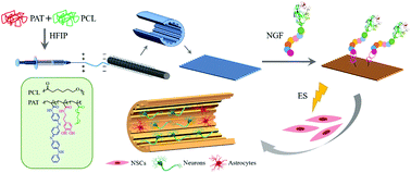
An effective treatment for spinal cord injury (SCI) remains a severe clinical challenge due to the intrinsically limited regenerative capacity and complex anatomical structure of the spinal cord. The combination of biomaterials, which serve as scaffolds for axonal growth, cells and neurotrophic factors, is an excellent candidate for spinal cord regeneration. Herein, a new micropatterned conductive electrospun nanofiber mesh was constructed with poly{[aniline tetramer methacrylamide]-co-[dopamine methacrylamide]-co-[poly(ethylene glycol) methyl ether methacrylate]}/PCL (PCAT) using a rotation electrospinning technology. The aim was to study the synergistic effects of electrical stimulation (ES) and a micropatterned conductive electrospun nanofiber mesh incorporated with nerve growth factor (NGF) on the differentiation of rat nerve stem cells (NSCs). The hydrophilicity of the conductive nanofiber mesh could be tailored by changing the dopamine (DA) and aniline tetramer (AT) content from 19° to 79°. A favorable electroactivity and conductivity was achieved by the AT segment of PCAT. The as-fabricated micropatterned electrospun nanofiber mesh possessed a regularly aligned valley and ridge structure, and the diameter of the nanofiber was 312 ± 58 nm, while the width of the valley and ridge was measured to be 210 ± 17 μm and 200 ± 16 μm, respectively. The growth and neurite outgrowth of differentiated NSCs were observed along the valley of the micropatterned nanofiber mesh. In addition, the NGF loaded micropatterned conductive electrospun nanofiber mesh combined with ES exhibited the highest cell viability, and effectively facilitated the differentiation of NSCs into neurons and suppressed the formation of astrocytes, thus exhibiting a great application potential for nerve tissue engineering.
中文翻译:

微图案导电电纺纳米纤维网与电刺激相结合,协同增强大鼠神经干细胞的分化
由于脊髓固有的再生能力和复杂的解剖结构,对脊髓损伤(SCI)的有效治疗仍然是严峻的临床挑战。用作轴突生长,细胞和神经营养因子的支架的生物材料的组合是脊髓再生的极佳候选者。在本文中,一个新的微图案化的导电电纺纳米纤维网用聚{[苯胺四聚体甲基丙烯酰胺]构建-共- [甲基丙烯酰胺多巴胺] -共-[聚乙二醇乙二醇甲基丙烯酸甲酯]} / PCL(PCAT)采用旋转电纺丝技术。目的是研究电刺激(ES)和掺有神经生长因子(NGF)的微图案导电电纺纳米纤维网对大鼠神经干细胞(NSCs)分化的协同作用。可以通过将多巴胺(DA)和苯胺四聚体(AT)的含量从19°更改为79°来调整导电纳米纤维网的亲水性。通过PCAT的AT段实现了良好的电活性和电导率。制成的微图案电纺纳米纤维网具有规则排列的谷脊结构,纳米纤维的直径为312±58 nm,而谷脊的宽度分别为210±17μm和200±16μm , 分别。沿微图案化纳米纤维网孔的山谷观察到分化的NSC的生长和神经突向外生长。此外,与ES结合的NGF负载微图案导电电纺纳米纤维网片具有最高的细胞活力,并有效地促进了NSCs向神经元的分化并抑制了星形胶质细胞的形成,因此在神经组织工程中具有巨大的应用潜力。
更新日期:2020-04-01
中文翻译:

微图案导电电纺纳米纤维网与电刺激相结合,协同增强大鼠神经干细胞的分化
由于脊髓固有的再生能力和复杂的解剖结构,对脊髓损伤(SCI)的有效治疗仍然是严峻的临床挑战。用作轴突生长,细胞和神经营养因子的支架的生物材料的组合是脊髓再生的极佳候选者。在本文中,一个新的微图案化的导电电纺纳米纤维网用聚{[苯胺四聚体甲基丙烯酰胺]构建-共- [甲基丙烯酰胺多巴胺] -共-[聚乙二醇乙二醇甲基丙烯酸甲酯]} / PCL(PCAT)采用旋转电纺丝技术。目的是研究电刺激(ES)和掺有神经生长因子(NGF)的微图案导电电纺纳米纤维网对大鼠神经干细胞(NSCs)分化的协同作用。可以通过将多巴胺(DA)和苯胺四聚体(AT)的含量从19°更改为79°来调整导电纳米纤维网的亲水性。通过PCAT的AT段实现了良好的电活性和电导率。制成的微图案电纺纳米纤维网具有规则排列的谷脊结构,纳米纤维的直径为312±58 nm,而谷脊的宽度分别为210±17μm和200±16μm , 分别。沿微图案化纳米纤维网孔的山谷观察到分化的NSC的生长和神经突向外生长。此外,与ES结合的NGF负载微图案导电电纺纳米纤维网片具有最高的细胞活力,并有效地促进了NSCs向神经元的分化并抑制了星形胶质细胞的形成,因此在神经组织工程中具有巨大的应用潜力。


 京公网安备 11010802027423号