Our official English website, www.x-mol.net, welcomes your
feedback! (Note: you will need to create a separate account there.)
Alterations in optical coherence tomography angiography findings in patients with high myopia
Eye ( IF 2.8 ) Pub Date : 2020-02-24 , DOI: 10.1038/s41433-020-0824-1 Turgay Ucak 1 , Erel Icel 1 , Hayati Yilmaz 1 , Yucel Karakurt 1 , Gamze Tasli 1 , Adem Ugurlu 1 , Erdinc Bozkurt 2
Eye ( IF 2.8 ) Pub Date : 2020-02-24 , DOI: 10.1038/s41433-020-0824-1 Turgay Ucak 1 , Erel Icel 1 , Hayati Yilmaz 1 , Yucel Karakurt 1 , Gamze Tasli 1 , Adem Ugurlu 1 , Erdinc Bozkurt 2
Affiliation
|
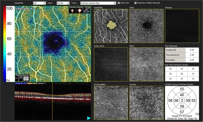
Purpose The aim of this study was to determine the macular changes using optical coherence tomography (OCT) and OCT-angiography (OCT-A) in eyes with high myopia. Determining the alterations in vascular structures can provide a clearer understanding of the pathophysiological mechanisms of this disease and help define new treatment options and preventive measures. Materials and methods Ninety-two patients with high myopia (axial length ≥ 26 mm) and 70 control cases without any known systemic or ocular diseases were enrolled in this prospective study. One eye of each patient was included in the statistical analyses. Results Retinal nerve fiber layer (RNFL) thickness and Early Treatment Diabetic Retinopathy Study (ETDRS) macula map values were lower in myopia compared with the controls. Both superior and inferior ganglion cell complex (GCC) thicknesses were significantly thinner in the high myopia compared with the controls ( p < 0.001). Regarding the OCT-A findings, although superficial or deep foveal avascular zones (FAZ) did not significantly differ between the two groups, the density values of superficial and deep microvessels were significantly lower in the high myopia group compared with the control cases. Conclusions In patients with high myopia, with an increase in the axial length and a decrease in RNFL and GCC thicknesses, the vascular densities of the superficial and deep retina were reduced in the macular region.
中文翻译:

高度近视患者光学相干断层扫描血管造影结果的变化
目的 本研究的目的是使用光学相干断层扫描 (OCT) 和 OCT 血管造影 (OCT-A) 确定高度近视眼的黄斑变化。确定血管结构的改变可以更清楚地了解这种疾病的病理生理机制,并有助于确定新的治疗方案和预防措施。材料和方法 92 名高度近视患者(轴长≥ 26 mm)和 70 名没有任何已知全身或眼部疾病的对照病例参加了这项前瞻性研究。每个患者的一只眼睛被包括在统计分析中。结果 与对照组相比,近视患者的视网膜神经纤维层 (RNFL) 厚度和早期治疗糖尿病视网膜病变研究 (ETDRS) 黄斑图值较低。与对照组相比,高度近视患者的上、下神经节细胞复合体 (GCC) 厚度均显着更薄 (p < 0.001)。关于OCT-A结果,虽然两组之间的浅表或深中央凹无血管区(FAZ)没有显着差异,但与对照组相比,高度近视组的表浅和深部微血管的密度值显着降低。结论 高度近视患者,随着眼轴长度的增加,RNFL和GCC厚度的减少,黄斑区视网膜浅层和深部的血管密度降低。尽管两组之间的浅表或深中央凹无血管区(FAZ)没有显着差异,但与对照组相比,高度近视组的表浅和深部微血管的密度值显着降低。结论 高度近视患者,随着眼轴长度的增加,RNFL和GCC厚度的减少,黄斑区视网膜浅层和深部的血管密度降低。尽管两组之间的浅表或深中央凹无血管区(FAZ)没有显着差异,但与对照组相比,高度近视组的表浅和深部微血管的密度值显着降低。结论 高度近视患者,随着眼轴长度的增加,RNFL和GCC厚度的减少,黄斑区视网膜浅层和深部的血管密度降低。
更新日期:2020-02-24
中文翻译:
高度近视患者光学相干断层扫描血管造影结果的变化
目的 本研究的目的是使用光学相干断层扫描 (OCT) 和 OCT 血管造影 (OCT-A) 确定高度近视眼的黄斑变化。确定血管结构的改变可以更清楚地了解这种疾病的病理生理机制,并有助于确定新的治疗方案和预防措施。材料和方法 92 名高度近视患者(轴长≥ 26 mm)和 70 名没有任何已知全身或眼部疾病的对照病例参加了这项前瞻性研究。每个患者的一只眼睛被包括在统计分析中。结果 与对照组相比,近视患者的视网膜神经纤维层 (RNFL) 厚度和早期治疗糖尿病视网膜病变研究 (ETDRS) 黄斑图值较低。与对照组相比,高度近视患者的上、下神经节细胞复合体 (GCC) 厚度均显着更薄 (p < 0.001)。关于OCT-A结果,虽然两组之间的浅表或深中央凹无血管区(FAZ)没有显着差异,但与对照组相比,高度近视组的表浅和深部微血管的密度值显着降低。结论 高度近视患者,随着眼轴长度的增加,RNFL和GCC厚度的减少,黄斑区视网膜浅层和深部的血管密度降低。尽管两组之间的浅表或深中央凹无血管区(FAZ)没有显着差异,但与对照组相比,高度近视组的表浅和深部微血管的密度值显着降低。结论 高度近视患者,随着眼轴长度的增加,RNFL和GCC厚度的减少,黄斑区视网膜浅层和深部的血管密度降低。尽管两组之间的浅表或深中央凹无血管区(FAZ)没有显着差异,但与对照组相比,高度近视组的表浅和深部微血管的密度值显着降低。结论 高度近视患者,随着眼轴长度的增加,RNFL和GCC厚度的减少,黄斑区视网膜浅层和深部的血管密度降低。


 京公网安备 11010802027423号