当前位置:
X-MOL 学术
›
Nat. Rev. Cardiol.
›
论文详情
Our official English website, www.x-mol.net, welcomes your
feedback! (Note: you will need to create a separate account there.)
Clinical quantitative cardiac imaging for the assessment of myocardial ischaemia.
Nature Reviews Cardiology ( IF 41.7 ) Pub Date : 2020-02-24 , DOI: 10.1038/s41569-020-0341-8 Marc Dewey 1, 2 , Maria Siebes 3 , Marc Kachelrieß 4 , Klaus F Kofoed 5 , Pál Maurovich-Horvat 6 , Konstantin Nikolaou 7 , Wenjia Bai 8 , Andreas Kofler 1 , Robert Manka 9, 10 , Sebastian Kozerke 10 , Amedeo Chiribiri 11 , Tobias Schaeffter 11, 12 , Florian Michallek 1 , Frank Bengel 13 , Stephan Nekolla 14 , Paul Knaapen 15 , Mark Lubberink 16, 17 , Roxy Senior 18 , Meng-Xing Tang 19 , Jan J Piek 20 , Tim van de Hoef 20 , Johannes Martens 21 , Laura Schreiber 21 ,
Nature Reviews Cardiology ( IF 41.7 ) Pub Date : 2020-02-24 , DOI: 10.1038/s41569-020-0341-8 Marc Dewey 1, 2 , Maria Siebes 3 , Marc Kachelrieß 4 , Klaus F Kofoed 5 , Pál Maurovich-Horvat 6 , Konstantin Nikolaou 7 , Wenjia Bai 8 , Andreas Kofler 1 , Robert Manka 9, 10 , Sebastian Kozerke 10 , Amedeo Chiribiri 11 , Tobias Schaeffter 11, 12 , Florian Michallek 1 , Frank Bengel 13 , Stephan Nekolla 14 , Paul Knaapen 15 , Mark Lubberink 16, 17 , Roxy Senior 18 , Meng-Xing Tang 19 , Jan J Piek 20 , Tim van de Hoef 20 , Johannes Martens 21 , Laura Schreiber 21 ,
Affiliation

|
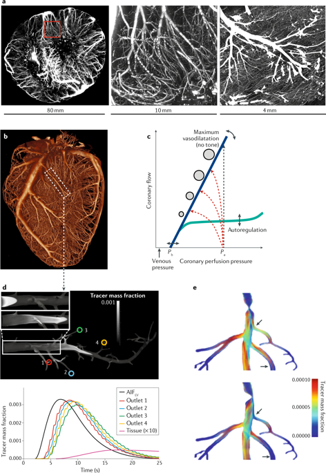
Cardiac imaging has a pivotal role in the prevention, diagnosis and treatment of ischaemic heart disease. SPECT is most commonly used for clinical myocardial perfusion imaging, whereas PET is the clinical reference standard for the quantification of myocardial perfusion. MRI does not involve exposure to ionizing radiation, similar to echocardiography, which can be performed at the bedside. CT perfusion imaging is not frequently used but CT offers coronary angiography data, and invasive catheter-based methods can measure coronary flow and pressure. Technical improvements to the quantification of pathophysiological parameters of myocardial ischaemia can be achieved. Clinical consensus recommendations on the appropriateness of each technique were derived following a European quantitative cardiac imaging meeting and using a real-time Delphi process. SPECT using new detectors allows the quantification of myocardial blood flow and is now also suited to patients with a high BMI. PET is well suited to patients with multivessel disease to confirm or exclude balanced ischaemia. MRI allows the evaluation of patients with complex disease who would benefit from imaging of function and fibrosis in addition to perfusion. Echocardiography remains the preferred technique for assessing ischaemia in bedside situations, whereas CT has the greatest value for combined quantification of stenosis and characterization of atherosclerosis in relation to myocardial ischaemia. In patients with a high probability of needing invasive treatment, invasive coronary flow and pressure measurement is well suited to guide treatment decisions. In this Consensus Statement, we summarize the strengths and weaknesses as well as the future technological potential of each imaging modality.
中文翻译:

临床定量心脏成像用于评估心肌缺血。
心脏成像在缺血性心脏病的预防,诊断和治疗中具有关键作用。SPECT最常用于临床心肌灌注显像,而PET是定量心肌灌注的临床参考标准。MRI不涉及暴露在电离辐射下,类似于超声心动图,可以在床边进行。CT灌注成像并不经常使用,但是CT提供冠状动脉造影数据,而基于侵入性导管的方法可以测量冠状动脉的流量和压力。可以实现对心肌缺血的病理生理参数定量的技术改进。在欧洲定量心脏成像会议之后,并使用实时Delphi过程得出了关于每种技术适用性的临床共识建议。使用新型检测器的SPECT可以量化心肌血流量,现在也适用于BMI高的患者。PET非常适合患有多支血管疾病的患者,以确认或排除局部缺血。MRI可以评估患有复杂疾病的患者,这些患者除了灌注外,还可以通过功能和纤维化的影像学获益。超声心动图仍然是评估床旁缺血的首选技术,而CT对于与心肌缺血相关的狭窄定量和动脉粥样硬化特征的组合量化具有最大的价值。对于极有可能需要进行侵入性治疗的患者,侵入性冠脉流量和压力测量非常适合指导治疗决策。在本共识声明中,
更新日期:2020-02-24
中文翻译:

临床定量心脏成像用于评估心肌缺血。
心脏成像在缺血性心脏病的预防,诊断和治疗中具有关键作用。SPECT最常用于临床心肌灌注显像,而PET是定量心肌灌注的临床参考标准。MRI不涉及暴露在电离辐射下,类似于超声心动图,可以在床边进行。CT灌注成像并不经常使用,但是CT提供冠状动脉造影数据,而基于侵入性导管的方法可以测量冠状动脉的流量和压力。可以实现对心肌缺血的病理生理参数定量的技术改进。在欧洲定量心脏成像会议之后,并使用实时Delphi过程得出了关于每种技术适用性的临床共识建议。使用新型检测器的SPECT可以量化心肌血流量,现在也适用于BMI高的患者。PET非常适合患有多支血管疾病的患者,以确认或排除局部缺血。MRI可以评估患有复杂疾病的患者,这些患者除了灌注外,还可以通过功能和纤维化的影像学获益。超声心动图仍然是评估床旁缺血的首选技术,而CT对于与心肌缺血相关的狭窄定量和动脉粥样硬化特征的组合量化具有最大的价值。对于极有可能需要进行侵入性治疗的患者,侵入性冠脉流量和压力测量非常适合指导治疗决策。在本共识声明中,


 京公网安备 11010802027423号