当前位置:
X-MOL 学术
›
Toxicol. Lett.
›
论文详情
Our official English website, www.x-mol.net, welcomes your
feedback! (Note: you will need to create a separate account there.)
Mass spectrometry imaging reveals lipid upregulation and bile acid changes indicating amitriptyline induced steatosis in a rat model
Toxicology Letters ( IF 2.9 ) Pub Date : 2020-06-01 , DOI: 10.1016/j.toxlet.2020.02.007 Judith M. Kampa , Mikail Sahin , Markus Slopianka , Marco Giampà , Hanna Bednarz , Rainer Ernst , Bjoern Riefke , Karsten Niehaus , Amol Fatangare
Toxicology Letters ( IF 2.9 ) Pub Date : 2020-06-01 , DOI: 10.1016/j.toxlet.2020.02.007 Judith M. Kampa , Mikail Sahin , Markus Slopianka , Marco Giampà , Hanna Bednarz , Rainer Ernst , Bjoern Riefke , Karsten Niehaus , Amol Fatangare
|
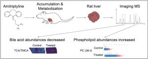
As a consequence of the detoxification process, drugs and drug related metabolites can accumulate in the liver, resulting in drug induced liver injury (DILI), which is the major cause for dose limitation. Amitriptyline, a commonly used tricyclic anti-depressant, is known to cause DILI. The mechanism of Amitriptyline induced liver injury is not yet completely understood. However, as it undergoes extensive hepatic metabolism, unraveling the molecular changes in the liver upon Amitriptyline treatment can help understand Amitriptyline's mode of toxicity. In this study, Amitriptyline treated male rat liver tissue was analyzed using Matrix Assisted Laser Desorption/Ionization-Mass Spectrometry Imaging (MALDI-MSI) to investigate the spatial abundances of Amitriptyline, lipids, and bile acids. The metabolism of Amitriptyline in liver tissue was successfully demonstrated, as the spatial distribution of Amitriptyline and its metabolites localize throughout treatment group liver samples. Several lipids appear upregulated, from which nine were identified as distinct phosphatidylcholine (PC) species. The detected bile acids were found to be lower in Amitriptyline treatment group. The combined results from histological findings, Oil Red O staining, and lipid zonation by MSI reveal lipid upregulation in the periportal area indicating drug induced macrovesicular steatosis (DIS).
中文翻译:

质谱成像显示脂质上调和胆汁酸变化表明阿米替林诱导大鼠模型脂肪变性
由于解毒过程,药物和药物相关代谢物会在肝脏中积聚,导致药物性肝损伤 (DILI),这是剂量限制的主要原因。阿米替林是一种常用的三环类抗抑郁药,已知会导致 DILI。阿米替林引起肝损伤的机制尚不完全清楚。然而,由于它经历了广泛的肝脏代谢,解开阿米替林治疗后肝脏的分子变化有助于了解阿米替林的毒性模式。在这项研究中,使用基质辅助激光解吸/电离质谱成像 (MALDI-MSI) 分析阿米替林处理的雄性大鼠肝组织,以研究阿米替林、脂质和胆汁酸的空间丰度。阿米替林在肝组织中的代谢得到了成功证明,因为阿米替林及其代谢物的空间分布在整个治疗组肝脏样本中都存在。几种脂质出现上调,其中九种被鉴定为不同的磷脂酰胆碱 (PC) 种类。发现检测到的胆汁酸在阿米替林治疗组中较低。来自组织学发现、油红 O 染色和 MSI 脂质分区的综合结果揭示了门静脉周围区域的脂质上调,表明药物诱导的大泡性脂肪变性 (DIS)。发现检测到的胆汁酸在阿米替林治疗组中较低。来自组织学发现、油红 O 染色和 MSI 脂质分区的综合结果揭示了门静脉周围区域的脂质上调,表明药物诱导的大泡性脂肪变性 (DIS)。发现检测到的胆汁酸在阿米替林治疗组中较低。来自组织学发现、油红 O 染色和 MSI 脂质分区的综合结果揭示了门静脉周围区域的脂质上调,表明药物诱导的大泡性脂肪变性 (DIS)。
更新日期:2020-06-01
中文翻译:
质谱成像显示脂质上调和胆汁酸变化表明阿米替林诱导大鼠模型脂肪变性
由于解毒过程,药物和药物相关代谢物会在肝脏中积聚,导致药物性肝损伤 (DILI),这是剂量限制的主要原因。阿米替林是一种常用的三环类抗抑郁药,已知会导致 DILI。阿米替林引起肝损伤的机制尚不完全清楚。然而,由于它经历了广泛的肝脏代谢,解开阿米替林治疗后肝脏的分子变化有助于了解阿米替林的毒性模式。在这项研究中,使用基质辅助激光解吸/电离质谱成像 (MALDI-MSI) 分析阿米替林处理的雄性大鼠肝组织,以研究阿米替林、脂质和胆汁酸的空间丰度。阿米替林在肝组织中的代谢得到了成功证明,因为阿米替林及其代谢物的空间分布在整个治疗组肝脏样本中都存在。几种脂质出现上调,其中九种被鉴定为不同的磷脂酰胆碱 (PC) 种类。发现检测到的胆汁酸在阿米替林治疗组中较低。来自组织学发现、油红 O 染色和 MSI 脂质分区的综合结果揭示了门静脉周围区域的脂质上调,表明药物诱导的大泡性脂肪变性 (DIS)。发现检测到的胆汁酸在阿米替林治疗组中较低。来自组织学发现、油红 O 染色和 MSI 脂质分区的综合结果揭示了门静脉周围区域的脂质上调,表明药物诱导的大泡性脂肪变性 (DIS)。发现检测到的胆汁酸在阿米替林治疗组中较低。来自组织学发现、油红 O 染色和 MSI 脂质分区的综合结果揭示了门静脉周围区域的脂质上调,表明药物诱导的大泡性脂肪变性 (DIS)。


 京公网安备 11010802027423号