Nature Medicine ( IF 58.7 ) Pub Date : 2020-02-17 , DOI: 10.1038/s41591-020-0770-2 Alvaro A Ordonez 1, 2, 3 , Hechuan Wang 4 , Gesham Magombedze 5 , Camilo A Ruiz-Bedoya 1, 2, 3 , Shashikant Srivastava 5 , Allen Chen 1, 6 , Elizabeth W Tucker 1, 2, 7 , Michael E Urbanowski 2, 8 , Lisa Pieterse 1, 2, 7 , E Fabian Cardozo 9 , Martin A Lodge 6 , Maunank R Shah 2, 8 , Daniel P Holt 6 , William B Mathews 6 , Robert F Dannals 6 , Jogarao V S Gobburu 4 , Charles A Peloquin 10 , Steven P Rowe 6 , Tawanda Gumbo 5 , Vijay D Ivaturi 4 , Sanjay K Jain 1, 2, 3, 6
|
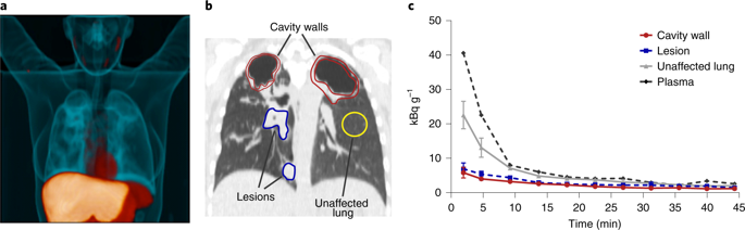
Tuberculosis (TB) is the leading cause of death from a single infectious agent, requiring at least 6 months of multidrug treatment to achieve cure1. However, the lack of reliable data on antimicrobial pharmacokinetics (PK) at infection sites hinders efforts to optimize antimicrobial dosing and shorten TB treatments2. In this study, we applied a new tool to perform unbiased, noninvasive and multicompartment measurements of antimicrobial concentration–time profiles in humans3. Newly identified patients with rifampin-susceptible pulmonary TB were enrolled in a first-in-human study4 using dynamic [11C]rifampin (administered as a microdose) positron emission tomography (PET) and computed tomography (CT). [11C]rifampin PET–CT was safe and demonstrated spatially compartmentalized rifampin exposures in pathologically distinct TB lesions within the same patients, with low cavity wall rifampin exposures. Repeat PET–CT measurements demonstrated independent temporal evolution of rifampin exposure trajectories in different lesions within the same patients. Similar findings were recapitulated by PET–CT in experimentally infected rabbits with cavitary TB and confirmed using postmortem mass spectrometry. Integrated modeling of the PET-captured concentration–time profiles in hollow-fiber bacterial kill curve experiments provided estimates on the rifampin dosing required to achieve cure in 4 months. These data, capturing the spatial and temporal heterogeneity of intralesional drug PK, have major implications for antimicrobial drug development.
中文翻译:
结核病患者的动态成像显示肺部病变中的异质药物暴露
结核病 (TB) 是导致单一传染性病原体死亡的主要原因,需要至少 6 个月的多种药物治疗才能治愈1。然而,在感染部位缺乏可靠的抗菌药代动力学 (PK) 数据,阻碍了优化抗菌药物剂量和缩短结核病治疗2的努力。在这项研究中,我们应用了一种新工具,对人体中的抗菌药物浓度-时间曲线进行无偏、无创和多室测量3。新发现的对利福平敏感的肺结核患者参加了一项首次人体研究4 ,该研究使用动态 [ 11 C] 利福平(作为微剂量给药)正电子发射断层扫描 (PET) 和计算机断层扫描 (CT)。[11C] 利福平 PET-CT 是安全的,并且在同一患者的病理不同的 TB 病灶中显示空间划分的利福平暴露,腔壁利福平暴露低。重复 PET-CT 测量表明,同一患者体内不同病变中利福平暴露轨迹的独立时间演变。PET-CT 在实验性感染的患有空洞性 TB 的兔子中概括了类似的发现,并使用死后质谱法证实了这一发现。中空纤维细菌杀灭曲线实验中 PET 捕获的浓度-时间曲线的综合建模提供了对 4 个月内实现治愈所需的利福平剂量的估计。这些数据捕获了病灶内药物 PK 的空间和时间异质性,对抗菌药物开发具有重要意义。


 京公网安备 11010802027423号