当前位置:
X-MOL 学术
›
JAMA Neurol.
›
论文详情
Our official English website, www.x-mol.net, welcomes your
feedback! (Note: you will need to create a separate account there.)
Temporal Dynamics of Cortical Microinfarcts in Cerebral Small Vessel Disease.
JAMA Neurology ( IF 20.4 ) Pub Date : 2020-05-01 , DOI: 10.1001/jamaneurol.2019.5106 Annemieke Ter Telgte 1 , Kim Wiegertjes 1 , Benno Gesierich 2 , Brendon Sri Baskaran 1 , José P Marques 3 , Hugo J Kuijf 4 , David G Norris 3 , Anil M Tuladhar 1 , Marco Duering 1, 2, 5 , Frank-Erik de Leeuw 1
JAMA Neurology ( IF 20.4 ) Pub Date : 2020-05-01 , DOI: 10.1001/jamaneurol.2019.5106 Annemieke Ter Telgte 1 , Kim Wiegertjes 1 , Benno Gesierich 2 , Brendon Sri Baskaran 1 , José P Marques 3 , Hugo J Kuijf 4 , David G Norris 3 , Anil M Tuladhar 1 , Marco Duering 1, 2, 5 , Frank-Erik de Leeuw 1
Affiliation

|
Importance
Neuropathology studies show a high prevalence of cortical microinfarcts (CMIs) in aging individuals, especially in patients with cerebrovascular disease and dementia. However, most, are invisible on T1- and T2-weighted magnetic resonance imaging (MRI), raising the question of how to explain this mismatch. Studies on small acute infarcts, detected on diffusion-weighted imaging (DWI), suggest that infarcts are largest in their acute phase and reduce in size thereafter. Therefore, we hypothesized that a subset of the CMI that are invisible on MRI can be detected on MRI in their acute phase. However, to our knowledge, a serial imaging study investigating the temporal dynamics of acute CMI (A-CMI) is lacking.
Objective
To determine the prevalence of chronic CMI (C-CMI) and the cumulative incidence and temporal dynamics of A-CMI in individuals with cerebral small vessel disease (SVD).
Design, Setting, Participants and Exposures
The RUN DMC-Intense study is a single-center hospital-based prospective cohort study on SVD performed between March 2016 and November 2017 and comprising 10 monthly 3-T MRI scans, including high-resolution DWI, 3-dimensional T1, 3-dimensional fluid-attenuated inversion recovery, and T2. One hundred six individuals from the previous longitudinal RUN DMC study were recruited based on the presence of progression of white matter hyperintensities on MRI between 2006 and 2015 and exclusion of causes of cerebral ischemia other than SVD. Fifty-four individuals (50.9%) participated. The median total follow-up duration was 39.5 weeks (interquartile range, 37.8-40.3). Statistical data analysis was performed between May and October 2019.
Main Outcomes and Measures
We determined the prevalence of C-CMI using the baseline T1, fluid-attenuated inversion recovery, and T2 scans. Monthly high-resolution DWI scans (n = 472) were screened to determine the cumulative incidence of A-CMI. The temporal dynamics of A-CMI were determined based on the MRI scans collected during the first follow-up visit after A-CMI onset and the last available follow-up visit.
Results
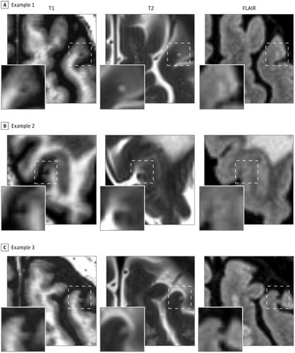
The median age of the cohort at baseline MRI was 69 years (interquartile range, 66-74 years) and 34 participants (63%) were men. The prevalence of C-CMI was 35% (95% CI, 0.24-0.49). Monthly DWI detected 21 A-CMI in 7 of 54 participants, resulting in a cumulative incidence of 13% (95% CI, 0.06-0.24). All A-CMI disappeared on follow-up MRI.
Conclusions and Relevance
Acute CMI never evolved into chronically MRI-detectable lesions. We suggest that these A-CMI underlie part of the submillimeter C-CMI encountered on neuropathological examination and thereby provide a source for the high CMI burden on neuropathology.
中文翻译:

大脑小血管疾病的皮质微梗塞的时间动态。
重要性神经病理学研究显示,老年人中,尤其是脑血管疾病和痴呆患者中,皮质微梗塞(CMI)患病率很高。但是,大多数在T1和T2加权磁共振成像(MRI)上不可见,这引发了如何解释这种失配的问题。对弥散加权成像(DWI)检测到的小急性梗死的研究表明,梗死在其急性期最大,随后缩小。因此,我们假设可以在急性期在MRI上检测到在MRI上不可见的CMI子集。然而,据我们所知,目前尚缺乏研究急性CMI(A-CMI)时空动态的连续影像学研究。目的确定脑小血管疾病(SVD)患者的慢性CMI(C-CMI)患病率以及A-CMI的累积发生率和时间动态。设计,设置,参与者和暴露RUN DMC-Intense研究是一项基于单中心医院的SVD前瞻性队列研究,于2016年3月至2017年11月进行,包括10个月的每月3T MRI扫描,包括高分辨率DWI,3维T1、3维流体衰减反演恢复和T2。根据2006年至2015年间MRI出现白质高信号的进展并排除了SVD以外的脑缺血的原因,招募了一百六十六名来自RUN DMC纵向研究的人。五十四人(50.9%)参加了会议。中位总随访时间为39岁。5周(四分位间距为37.8-40.3)。在2019年5月至2019年10月之间进行了统计数据分析。主要结果和措施我们使用基线T1,液体衰减反转恢复和T2扫描确定了C-CMI的患病率。每月进行高分辨率DWI扫描(n = 472),以确定A-CMI的累积发生率。A-CMI的时空动态是根据A-CMI发作后的首次随访和最后一次随访中收集的MRI扫描确定的。结果基线MRI队列的中位年龄为69岁(四分位间距为66-74岁),其中34位参与者(63%)为男性。C-CMI的患病率为35%(95%CI,0.24-0.49)。每月DWI在54位参与者中的7位中检测到21位A-CMI,累计发生率为13%(95%CI,0.06-0.24)。所有A-CMI在随访MRI中均消失。结论和相关性急性CMI从未演变成可在MRI上检测到的慢性病变。我们建议这些A-CMI是在神经病理学检查中遇到的亚毫米C-CMI的一部分,从而为神经病理学的高CMI负担提供了来源。
更新日期:2020-05-01
中文翻译:

大脑小血管疾病的皮质微梗塞的时间动态。
重要性神经病理学研究显示,老年人中,尤其是脑血管疾病和痴呆患者中,皮质微梗塞(CMI)患病率很高。但是,大多数在T1和T2加权磁共振成像(MRI)上不可见,这引发了如何解释这种失配的问题。对弥散加权成像(DWI)检测到的小急性梗死的研究表明,梗死在其急性期最大,随后缩小。因此,我们假设可以在急性期在MRI上检测到在MRI上不可见的CMI子集。然而,据我们所知,目前尚缺乏研究急性CMI(A-CMI)时空动态的连续影像学研究。目的确定脑小血管疾病(SVD)患者的慢性CMI(C-CMI)患病率以及A-CMI的累积发生率和时间动态。设计,设置,参与者和暴露RUN DMC-Intense研究是一项基于单中心医院的SVD前瞻性队列研究,于2016年3月至2017年11月进行,包括10个月的每月3T MRI扫描,包括高分辨率DWI,3维T1、3维流体衰减反演恢复和T2。根据2006年至2015年间MRI出现白质高信号的进展并排除了SVD以外的脑缺血的原因,招募了一百六十六名来自RUN DMC纵向研究的人。五十四人(50.9%)参加了会议。中位总随访时间为39岁。5周(四分位间距为37.8-40.3)。在2019年5月至2019年10月之间进行了统计数据分析。主要结果和措施我们使用基线T1,液体衰减反转恢复和T2扫描确定了C-CMI的患病率。每月进行高分辨率DWI扫描(n = 472),以确定A-CMI的累积发生率。A-CMI的时空动态是根据A-CMI发作后的首次随访和最后一次随访中收集的MRI扫描确定的。结果基线MRI队列的中位年龄为69岁(四分位间距为66-74岁),其中34位参与者(63%)为男性。C-CMI的患病率为35%(95%CI,0.24-0.49)。每月DWI在54位参与者中的7位中检测到21位A-CMI,累计发生率为13%(95%CI,0.06-0.24)。所有A-CMI在随访MRI中均消失。结论和相关性急性CMI从未演变成可在MRI上检测到的慢性病变。我们建议这些A-CMI是在神经病理学检查中遇到的亚毫米C-CMI的一部分,从而为神经病理学的高CMI负担提供了来源。


 京公网安备 11010802027423号