Our official English website, www.x-mol.net, welcomes your feedback! (Note: you will need to create a separate account there.)
MultiColor imaging to detect different subtypes of retinal microaneurysms in diabetic retinopathy
Eye ( IF 3.9 ) Pub Date : 2020-02-17 , DOI: 10.1038/s41433-020-0811-6 Alessandro Arrigo 1 , Michel Teussink 2 , Emanuela Aragona 1 , Francesco Bandello 1 , Maurizio Battaglia Parodi 1
Eye ( IF 3.9 ) Pub Date : 2020-02-17 , DOI: 10.1038/s41433-020-0811-6 Alessandro Arrigo 1 , Michel Teussink 2 , Emanuela Aragona 1 , Francesco Bandello 1 , Maurizio Battaglia Parodi 1
Affiliation
|
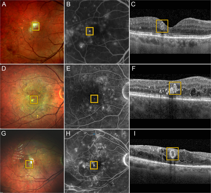
Background Retinal microaneurysms (MAs) are among the earliest signs of diabetic retinopathy (DR) and are typically detected by fluorescein angiography (FA). Confocal MultiColor is a noninvasive-imaging technique able to analyze different retinal features by capturing three simultaneous reflectance images. The main aim of the present study was to characterize morphological features of MAs by means of MultiColor images and to compare these with spectral domain optical coherence tomography (SD-OCT) and FA findings. Methods A cross-sectional, observational study setting was chosen. Multimodal imaging included MultiColor, SD-OCT and FA images. We performed a qualitative analysis in order to assess the relationship between MultiColor and its green- and red-reflectance components, SD-OCT (hyperreflective, hyporeflective and mixed reflectivity) and FA findings. MAs detected on our MultiColor images were then categorized in accordance with a previously published histological classification. Results In our study FA images were used to detect 153 MAs in 30 eyes displaying DR. MultiColor was able to distinguish 122 MAs (80%). We identified green (16%), red (19%), and mixed (65%) MAs, corresponding to different reflectivity features detected by SD-OCT. MAs not visualized on MultiColor images corresponded to tiny hyperreflective lesions on SD-OCT. We compared our imaging findings with a histological MA classification reported in the literature. Our findings showed a strict relationship between MA subtypes and SD-OCT, suggesting that the composition of MAs (cells + endothelium + fibrosis) may influence the signal detected in MultiColor images. Conclusions MultiColor appears to be a useful technique for investigating MA features in patients with DR.
中文翻译:

多色成像检测糖尿病视网膜病变中不同亚型的视网膜微动脉瘤
背景 视网膜微动脉瘤 (MA) 是糖尿病视网膜病变 (DR) 的最早征兆之一,通常通过荧光素血管造影 (FA) 检测到。共聚焦多色是一种无创成像技术,能够通过同时捕获三个反射图像来分析不同的视网膜特征。本研究的主要目的是通过多色图像表征 MA 的形态特征,并将其与光谱域光学相干断层扫描 (SD-OCT) 和 FA 发现进行比较。方法 选择横断面观察性研究设置。多模态成像包括多色、SD-OCT 和 FA 图像。我们进行了定性分析,以评估 MultiColor 与其绿色和红色反射分量 SD-OCT(超反射、低反射率和混合反射率)和 FA 结果。然后根据先前公布的组织学分类对我们的多色图像上检测到的 MA 进行分类。结果在我们的研究中,FA 图像用于检测 30 只显示 DR 的眼睛中的 153 个 MA。MultiColor 能够区分 122 个 MA (80%)。我们确定了绿色 (16%)、红色 (19%) 和混合 (65%) MA,对应于 SD-OCT 检测到的不同反射率特征。在多色图像上未显示的 MA 对应于 SD-OCT 上的微小超反射病变。我们将我们的影像学发现与文献中报道的组织学 MA 分类进行了比较。我们的研究结果显示 MA 亚型与 SD-OCT 之间存在严格关系,这表明 MA 的组成(细胞 + 内皮 + 纤维化)可能会影响多色图像中检测到的信号。
更新日期:2020-02-17
中文翻译:
多色成像检测糖尿病视网膜病变中不同亚型的视网膜微动脉瘤
背景 视网膜微动脉瘤 (MA) 是糖尿病视网膜病变 (DR) 的最早征兆之一,通常通过荧光素血管造影 (FA) 检测到。共聚焦多色是一种无创成像技术,能够通过同时捕获三个反射图像来分析不同的视网膜特征。本研究的主要目的是通过多色图像表征 MA 的形态特征,并将其与光谱域光学相干断层扫描 (SD-OCT) 和 FA 发现进行比较。方法 选择横断面观察性研究设置。多模态成像包括多色、SD-OCT 和 FA 图像。我们进行了定性分析,以评估 MultiColor 与其绿色和红色反射分量 SD-OCT(超反射、低反射率和混合反射率)和 FA 结果。然后根据先前公布的组织学分类对我们的多色图像上检测到的 MA 进行分类。结果在我们的研究中,FA 图像用于检测 30 只显示 DR 的眼睛中的 153 个 MA。MultiColor 能够区分 122 个 MA (80%)。我们确定了绿色 (16%)、红色 (19%) 和混合 (65%) MA,对应于 SD-OCT 检测到的不同反射率特征。在多色图像上未显示的 MA 对应于 SD-OCT 上的微小超反射病变。我们将我们的影像学发现与文献中报道的组织学 MA 分类进行了比较。我们的研究结果显示 MA 亚型与 SD-OCT 之间存在严格关系,这表明 MA 的组成(细胞 + 内皮 + 纤维化)可能会影响多色图像中检测到的信号。


 京公网安备 11010802027423号