Our official English website, www.x-mol.net, welcomes your
feedback! (Note: you will need to create a separate account there.)
Widefield topographical analysis of the retinal perfusion and neuroretinal thickness in healthy eyes: a pilot study
Eye ( IF 2.8 ) Pub Date : 2020-02-13 , DOI: 10.1038/s41433-020-0804-5 Enrico Borrelli 1 , Lisa Toto 1 , Pasquale Viggiano 1 , Federica Evangelista 1 , Michele Palmieri 1 , Rodolfo Mastropasqua 2
Eye ( IF 2.8 ) Pub Date : 2020-02-13 , DOI: 10.1038/s41433-020-0804-5 Enrico Borrelli 1 , Lisa Toto 1 , Pasquale Viggiano 1 , Federica Evangelista 1 , Michele Palmieri 1 , Rodolfo Mastropasqua 2
Affiliation
|
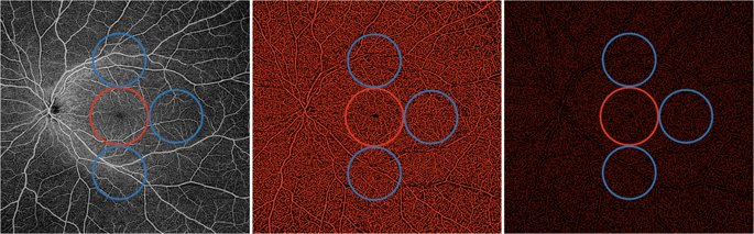
Purpose In this pilot study we reported variation of superficial (SCP) and deep (DCP) capillary plexuses flow in macular and near/mid periphery regions in healthy subjects using widefield swept source-optical coherence tomography angiography (SS-OCTA). Methods In this prospective, cross-sectional study, enroled subjects were imaged with an SS-OCTA system (PLEX Elite 9000, Carl Zeiss Meditec Inc., Dublin, CA, USA). OCTA scans were taken in primary and extremes of gaze and a montage was automatically created. Quantitative analysis was performed in the macular and peripheral regions. In addition, SCP and DCP variables were further investigated in distinct fields within these three different regions. Results Fifty-five young healthy subjects (55 eyes) were enroled. The retinal periphery displayed a higher SCP perfusion density (39.6 ± 1.7% and 40.7 ± 1.4%, P < 0.0001) and SCP vessel diameter index (3.5 ± 0.2 and 3.6 ± 0.2, P < 0.0001), in comparison with the macular region. At the DCP level, the retinal periphery was characterized by a lower perfusion density (41.6 ± 3.7% and 37.9 ± 2.9%, P < 0.0001) and vessel length density (14.6 ± 6.0% and 9.9 ± 2.6%, P < 0.0001). In the analysis investigating the DCP in the retinal periphery, the temporal sector was characterized by a reduction in perfusion density, vessel length density, and vessel diameter index. In univariate analysis, the retinal thickness was found to have a significant direct relationship with DCP perfusion density ( P < 0.0001), but not with SCP perfusion density ( P = 0.712). Conclusions We report quantitative mapping of the SCP and DCP in healthy individuals. The DCP perfusion appears to have a wide topographical variation, which is strictly dependent on the retinal thickness.
中文翻译:

健康眼视网膜灌注和神经视网膜厚度的宽场地形分析:一项试点研究
目的 在这项初步研究中,我们使用宽场扫描源光学相干断层扫描血管造影 (SS-OCTA) 报告了健康受试者黄斑和近/中周边区域浅层 (SCP) 和深层 (DCP) 毛细血管丛血流的变化。方法 在这项前瞻性横断面研究中,使用 SS-OCTA 系统(PLEX Elite 9000,Carl Zeiss Meditec Inc.,都柏林,加利福尼亚州,美国)对入选受试者进行成像。OCTA 扫描是在初级和极端注视下进行的,并自动创建蒙太奇。在黄斑和周边区域进行定量分析。此外,SCP 和 DCP 变量在这三个不同区域的不同领域进行了进一步研究。结果 纳入 55 名年轻健康受试者(55 只眼)。与黄斑区相比,视网膜周边显示出更高的 SCP 灌注密度(39.6 ± 1.7% 和 40.7 ± 1.4%,P < 0.0001)和 SCP 血管直径指数(3.5 ± 0.2 和 3.6 ± 0.2,P < 0.0001)。在DCP水平,视网膜周边的特点是较低的灌注密度(41.6±3.7%和37.9±2.9%,P<0.0001)和血管长度密度(14.6±6.0%和9.9±2.6%,P<0.0001)。在研究视网膜周边 DCP 的分析中,颞区的特点是灌注密度、血管长度密度和血管直径指数降低。在单变量分析中,发现视网膜厚度与DCP灌注密度有显着的直接关系(P<0.0001),但与SCP灌注密度没有显着的直接关系(P=0.712)。结论 我们报告了健康个体中 SCP 和 DCP 的定量图谱。DCP 灌注似乎具有广泛的地形变化,这严格取决于视网膜厚度。
更新日期:2020-02-13
中文翻译:
健康眼视网膜灌注和神经视网膜厚度的宽场地形分析:一项试点研究
目的 在这项初步研究中,我们使用宽场扫描源光学相干断层扫描血管造影 (SS-OCTA) 报告了健康受试者黄斑和近/中周边区域浅层 (SCP) 和深层 (DCP) 毛细血管丛血流的变化。方法 在这项前瞻性横断面研究中,使用 SS-OCTA 系统(PLEX Elite 9000,Carl Zeiss Meditec Inc.,都柏林,加利福尼亚州,美国)对入选受试者进行成像。OCTA 扫描是在初级和极端注视下进行的,并自动创建蒙太奇。在黄斑和周边区域进行定量分析。此外,SCP 和 DCP 变量在这三个不同区域的不同领域进行了进一步研究。结果 纳入 55 名年轻健康受试者(55 只眼)。与黄斑区相比,视网膜周边显示出更高的 SCP 灌注密度(39.6 ± 1.7% 和 40.7 ± 1.4%,P < 0.0001)和 SCP 血管直径指数(3.5 ± 0.2 和 3.6 ± 0.2,P < 0.0001)。在DCP水平,视网膜周边的特点是较低的灌注密度(41.6±3.7%和37.9±2.9%,P<0.0001)和血管长度密度(14.6±6.0%和9.9±2.6%,P<0.0001)。在研究视网膜周边 DCP 的分析中,颞区的特点是灌注密度、血管长度密度和血管直径指数降低。在单变量分析中,发现视网膜厚度与DCP灌注密度有显着的直接关系(P<0.0001),但与SCP灌注密度没有显着的直接关系(P=0.712)。结论 我们报告了健康个体中 SCP 和 DCP 的定量图谱。DCP 灌注似乎具有广泛的地形变化,这严格取决于视网膜厚度。


 京公网安备 11010802027423号