Our official English website, www.x-mol.net, welcomes your
feedback! (Note: you will need to create a separate account there.)
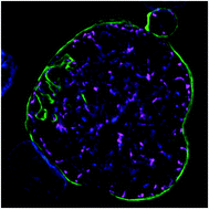
An adaptable soft-mold embossing process for fabricating optically-accessible, microfeature-based culture systems and application toward liver stage antimalarial compound testing.
Lab on a Chip ( IF 6.1 ) Pub Date : 2020-02-14 , DOI: 10.1039/c9lc00921c Steven P Maher 1 , Amy J Conway 2 , Alison Roth 2 , Swamy R Adapa 2 , Phillip Cualing 2 , Chiara Andolina 3 , James Hsiao 4 , Jessica Turgeon 2 , Victor Chaumeau 3 , Myles Johnson 2 , Chris Palmiotti 4 , Naresh Singh 2 , Samantha J Barnes 2 , Raahil Patel 2 , Virginia Van Grod 4 , Robert Carter 2 , H-C Steve Sun 4 , Jetsumon Sattabongkot 5 , Brice Campo 6 , François Nosten 3 , Wajeeh M Saadi 4 , John H Adams 2 , Rays H Y Jiang 2 , Dennis E Kyle 1
Lab on a Chip ( IF 6.1 ) Pub Date : 2020-02-14 , DOI: 10.1039/c9lc00921c Steven P Maher 1 , Amy J Conway 2 , Alison Roth 2 , Swamy R Adapa 2 , Phillip Cualing 2 , Chiara Andolina 3 , James Hsiao 4 , Jessica Turgeon 2 , Victor Chaumeau 3 , Myles Johnson 2 , Chris Palmiotti 4 , Naresh Singh 2 , Samantha J Barnes 2 , Raahil Patel 2 , Virginia Van Grod 4 , Robert Carter 2 , H-C Steve Sun 4 , Jetsumon Sattabongkot 5 , Brice Campo 6 , François Nosten 3 , Wajeeh M Saadi 4 , John H Adams 2 , Rays H Y Jiang 2 , Dennis E Kyle 1
Affiliation
|
Advanced cell culture methods for modeling organ-level structure have been demonstrated to replicate in vivo conditions more accurately than traditional in vitro cell culture. Given that the liver is particularly important to human health, several advanced culture methods have been developed to experiment with liver disease states, including infection with Plasmodium parasites, the causative agent of malaria. These models have demonstrated that intrahepatic parasites require functionally stable hepatocytes to thrive and robust characterization of the parasite populations' response to investigational therapies is dependent on high-content and high-resolution imaging (HC/RI). We previously reported abiotic confinement extends the functional longevity of primary hepatocytes in a microfluidic platform and set out to instill confinement in a microtiter plate platform while maintaining optical accessibility for HC/RI; with an end-goal of producing an improved P. vivax liver stage culture model. We developed a novel fabrication process in which a PDMS soft mold embosses hepatocyte-confining microfeatures into polystyrene, resulting in microfeature-based hepatocyte confinement (μHEP) slides and plates. Our process was optimized to form both microfeatures and culture wells in a single embossing step, resulting in a 100 μm-thick bottom ideal for HC/RI, and was found inexpensively amendable to microfeature design changes. Microfeatures improved intrahepatic parasite infection rates and μHEP systems were used to reconfirm the activity of reference antimalarials in phenotypic dose-response assays. RNAseq of hepatocytes in μHEP systems demonstrated microfeatures sustain hepatic differentiation and function, suggesting broader utility for preclinical hepatic assays; while our tailorable embossing process could be repurposed for developing additional organ models.
中文翻译:

一种适应性强的软模压花工艺,用于制造光学可访问的,基于微特征的培养系统,并应用于肝阶段抗疟化合物测试。
与传统的体外细胞培养相比,已证明用于建模器官水平结构的先进细胞培养方法可以更准确地复制体内条件。鉴于肝脏对人类健康特别重要,因此已经开发出了几种先进的培养方法来试验肝脏疾病,包括感染疟原虫的疟原虫寄生虫。这些模型表明,肝内寄生虫需要功能稳定的肝细胞才能蓬勃发展,并且寄生虫种群对研究疗法的反应的强劲表征取决于高内涵和高分辨率成像(HC / RI)。我们先前曾报道非生物限制在微流控平台上延长了原代肝细胞的功能寿命,并开始将其滴注在微量滴定板平台上,同时保持了HC / RI的光学可及性。最终产生了改良的间日疟原虫肝阶段培养模型。我们开发了一种新颖的制造工艺,其中PDMS软模将含肝细胞的微特征压印到聚苯乙烯中,从而导致基于微特征的肝细胞限制(μHEP)玻片和板。我们的工艺经过优化,可在单个压印步骤中同时形成微特征和培养孔,从而使HC / RI的底部底部厚度为100μm,并且发现微特征设计变更可廉价地进行修改。微特征提高了肝内寄生虫感染率,μHEP系统用于在表型剂量反应分析中确认参考抗疟药的活性。在μHEP系统中,肝细胞的RNAseq显示出微特征可维持肝的分化和功能,这表明其在临床前肝试验中的应用更为广泛。同时我们可定制的压花工艺可用于开发其他器官模型。
更新日期:2020-03-19
中文翻译:
一种适应性强的软模压花工艺,用于制造光学可访问的,基于微特征的培养系统,并应用于肝阶段抗疟化合物测试。
与传统的体外细胞培养相比,已证明用于建模器官水平结构的先进细胞培养方法可以更准确地复制体内条件。鉴于肝脏对人类健康特别重要,因此已经开发出了几种先进的培养方法来试验肝脏疾病,包括感染疟原虫的疟原虫寄生虫。这些模型表明,肝内寄生虫需要功能稳定的肝细胞才能蓬勃发展,并且寄生虫种群对研究疗法的反应的强劲表征取决于高内涵和高分辨率成像(HC / RI)。我们先前曾报道非生物限制在微流控平台上延长了原代肝细胞的功能寿命,并开始将其滴注在微量滴定板平台上,同时保持了HC / RI的光学可及性。最终产生了改良的间日疟原虫肝阶段培养模型。我们开发了一种新颖的制造工艺,其中PDMS软模将含肝细胞的微特征压印到聚苯乙烯中,从而导致基于微特征的肝细胞限制(μHEP)玻片和板。我们的工艺经过优化,可在单个压印步骤中同时形成微特征和培养孔,从而使HC / RI的底部底部厚度为100μm,并且发现微特征设计变更可廉价地进行修改。微特征提高了肝内寄生虫感染率,μHEP系统用于在表型剂量反应分析中确认参考抗疟药的活性。在μHEP系统中,肝细胞的RNAseq显示出微特征可维持肝的分化和功能,这表明其在临床前肝试验中的应用更为广泛。同时我们可定制的压花工艺可用于开发其他器官模型。


 京公网安备 11010802027423号