当前位置:
X-MOL 学术
›
ACS Chem. Neurosci.
›
论文详情
Our official English website, www.x-mol.net, welcomes your
feedback! (Note: you will need to create a separate account there.)
Enhanced Mitophagy Activity in Prion-Infected Cultured Cells and Prion-Infected Experimental Mice via a Pink1/Parkin-Dependent Mitophagy Pathway.
ACS Chemical Neuroscience ( IF 4.1 ) Pub Date : 2020-02-25 , DOI: 10.1021/acschemneuro.0c00039 Li-Ping Gao 1 , Kang Xiao 1 , Yue-Zhang Wu 1 , Dong-Dong Chen 1 , Xue-Hua Yang 1 , Qi Shi 1, 2 , Xiao-Ping Dong 1, 2, 3, 4
ACS Chemical Neuroscience ( IF 4.1 ) Pub Date : 2020-02-25 , DOI: 10.1021/acschemneuro.0c00039 Li-Ping Gao 1 , Kang Xiao 1 , Yue-Zhang Wu 1 , Dong-Dong Chen 1 , Xue-Hua Yang 1 , Qi Shi 1, 2 , Xiao-Ping Dong 1, 2, 3, 4
Affiliation
|
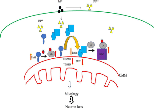
Mitophagy is an important process for removing damaged mitochondria in cells, the dysfunction of which has been directly linked to an increasing number of neurodegenerative disorders. However, the details of mitophagy in prion diseases still need to be deeply explored. In this study, we identified more autophagosomes and large swelling mitochondria structures in the prion-infected cultured cell line SMB-S15 by transmission electron microscopy, accompanying the molecular evidence of activated autophagic flux. Western blots illustrated that the levels of Pink1 and Parkin, particularly in the mitochondrial fraction, were increased in SMB-S15 cells, whereas the levels of mitochondrial membrane proteins TIMM44, TOMM20, and TIMM23 were decreased. The amount of whole polyubiquitinated proteins decreased, but that of phosphor-polyubiquitinated proteins increased in SMB-S15 cells. The level of MFN2 in SMB-S15 cells were down-regulated, but its polyubiquitinated form was up-regulated. Knockdown of the expressions of Pink1 and Parkin by the individual SiRNAs in SMB-S15 cells reduced autophagic activity but did not seem to influence the expressions of TOMM20 and TIMM23. Moreover, we also demonstrated that the brain levels of Pink1 and Parkin in the mice infected with scrapie strains 139A and ME7 were remarkably increased at the terminal stage of the disease by Western blot and immunohistochemical (IHC) assays. Immunofluorescent assays revealed that Pink1 signals widely colocalized with GAFP-, Iba1-, and NeuN-positive cells in the brains of scrapie-infected mice. IHC assays with serial sections of the brain tissues infected with agents 139A and ME7 showed more Pink1- and Parkin-positive cells located at the areas with more PrPSc deposit. These results suggest an activated mitophagy in prion-infected cells and prion-infected experimental mice, probably via an enhanced Pink-Parkin pathway.
中文翻译:

通过Pink1 / Parkin依赖线粒体途径在Pri病毒感染的培养细胞和Pri病毒感染的实验小鼠中增强线粒体活性。
线粒体吞噬是去除细胞中受损线粒体的重要过程,线粒体的功能障碍与越来越多的神经退行性疾病直接相关。然而,病毒疾病中线粒体的细节仍需深入探讨。在这项研究中,我们通过透射电子显微镜在identified病毒感染的培养细胞系SMB-S15中鉴定了更多的自噬体和大肿胀的线粒体结构,并伴有激活的自噬通量的分子证据。Western印迹表明,SMB-S15细胞中Pink1和Parkin的水平,尤其是线粒体部分的水平增加,而线粒体膜蛋白TIMM44,TOMM20和TIMM23的水平降低。整个多泛素化蛋白的量减少了,但是在SMB-S15细胞中,磷酸化多泛素化蛋白的含量增加。SMB-S15细胞中MFN2的水平被下调,但其多泛素化形式被上调。SMB-S15细胞中的单个SiRNA抑制Pink1和Parkin的表达降低了自噬活性,但似乎并未影响TOMM20和TIMM23的表达。此外,我们还证明了通过Western blot和免疫组化(IHC)分析,在疾病终末期,感染了瘙痒病菌株139A和ME7的小鼠的Pink1和Parkin的脑水平显着增加。免疫荧光分析表明,Pink1信号在被瘙痒病感染的小鼠的大脑中与GAFP,Iba1和NeuN阳性细胞广泛共定位。用感染了139A和ME7的脑组织的连续切片进行的IHC分析显示,更多的Pink1-和Parkin阳性细胞位于具有更多PrPSc沉积物的区域。这些结果表明in病毒感染的细胞和病毒感染的实验小鼠可能通过增强的粉红色-帕金途径激活了线粒体。
更新日期:2020-02-26
中文翻译:
通过Pink1 / Parkin依赖线粒体途径在Pri病毒感染的培养细胞和Pri病毒感染的实验小鼠中增强线粒体活性。
线粒体吞噬是去除细胞中受损线粒体的重要过程,线粒体的功能障碍与越来越多的神经退行性疾病直接相关。然而,病毒疾病中线粒体的细节仍需深入探讨。在这项研究中,我们通过透射电子显微镜在identified病毒感染的培养细胞系SMB-S15中鉴定了更多的自噬体和大肿胀的线粒体结构,并伴有激活的自噬通量的分子证据。Western印迹表明,SMB-S15细胞中Pink1和Parkin的水平,尤其是线粒体部分的水平增加,而线粒体膜蛋白TIMM44,TOMM20和TIMM23的水平降低。整个多泛素化蛋白的量减少了,但是在SMB-S15细胞中,磷酸化多泛素化蛋白的含量增加。SMB-S15细胞中MFN2的水平被下调,但其多泛素化形式被上调。SMB-S15细胞中的单个SiRNA抑制Pink1和Parkin的表达降低了自噬活性,但似乎并未影响TOMM20和TIMM23的表达。此外,我们还证明了通过Western blot和免疫组化(IHC)分析,在疾病终末期,感染了瘙痒病菌株139A和ME7的小鼠的Pink1和Parkin的脑水平显着增加。免疫荧光分析表明,Pink1信号在被瘙痒病感染的小鼠的大脑中与GAFP,Iba1和NeuN阳性细胞广泛共定位。用感染了139A和ME7的脑组织的连续切片进行的IHC分析显示,更多的Pink1-和Parkin阳性细胞位于具有更多PrPSc沉积物的区域。这些结果表明in病毒感染的细胞和病毒感染的实验小鼠可能通过增强的粉红色-帕金途径激活了线粒体。


 京公网安备 11010802027423号