当前位置:
X-MOL 学术
›
NMR Biomed.
›
论文详情
Our official English website, www.x-mol.net, welcomes your
feedback! (Note: you will need to create a separate account there.)
The capability of detecting small vessels beyond the conventional MRI sensitivity using iron-based contrast agent enhanced susceptibility weighted imaging.
NMR in Biomedicine ( IF 2.7 ) Pub Date : 2020-02-11 , DOI: 10.1002/nbm.4256 Haoyu Wang 1 , Quan Jiang 2 , Yimin Shen 3 , Li Zhang 2 , E Mark Haacke 3 , Yulin Ge 4 , Shouliang Qi 5 , Jiani Hu 3
NMR in Biomedicine ( IF 2.7 ) Pub Date : 2020-02-11 , DOI: 10.1002/nbm.4256 Haoyu Wang 1 , Quan Jiang 2 , Yimin Shen 3 , Li Zhang 2 , E Mark Haacke 3 , Yulin Ge 4 , Shouliang Qi 5 , Jiani Hu 3
Affiliation
|
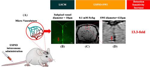
Imaging brain microvasculature is important in cerebrovascular diseases. However, there is still a lack of non-invasive, non-radiation, and whole-body imaging techniques to investigate them. The aim of this study is to develop an ultra-small superparamagnetic iron oxide (USPIO) enhanced susceptibility weighted imaging (SWI) method for imaging micro-vasculature in both animal (~10 μm in rat) and human brain. We hypothesized that the USPIO-SWI technique could improve the detection sensitivity of the diameter of small subpixel vessels 10-fold compared with conventional MRI methods. Computer simulations were first performed with a double-cylinder digital model to investigate the theoretical basis for this hypothesis. The theoretical results were verified using in vitro phantom studies and in vivo rat MRI studies (n = 6) with corresponding ex vivo histological examinations. Additionally, in vivo human studies (n = 3) were carried out to demonstrate the translational power of the USPIO-SWI method. By directly comparing the small vessel diameters of an in vivo rat using USPIO-SWI with the small vessel diameters of the corresponding histological slide using laser scanning confocal microscopy, 13.3-fold and 19.9-fold increases in SWI apparent diameter were obtained with 5.6 mg Fe/kg and 16.8 mg Fe/kg ferumoxytol, respectively. The USPIO-SWI method exhibited its excellent ability to detect small vessels down to about 10 μm diameter in rat brain. The in vivo human study unveiled hidden arterioles and venules and demonstrated its potential in clinical practice. Theoretical modeling simulations and in vitro phantom studies also confirmed a more than 10-fold increase in the USPIO-SWI apparent diameter compared with the actual small vessel diameter size. It is feasible to use SWI blooming effects induced by USPIO to detect small vessels (down to 10 μm in diameter for rat brain), well beyond the spatial resolution limit of conventional MRI methods. The USPIO-SWI method demonstrates higher potential in cerebrovascular disease investigations.
中文翻译:

使用铁基造影剂检测常规MRI灵敏度以外的小血管的能力增强了磁化率加权成像。
脑微血管成像对脑血管疾病很重要。但是,仍然缺乏非侵入性,非辐射和全身成像技术来进行研究。这项研究的目的是开发一种超小型超顺磁性氧化铁(USPIO)增强磁化率加权成像(SWI)方法,以对动物(大鼠约10μm)和人脑中的微血管成像。我们假设,与传统的MRI方法相比,USPIO-SWI技术可以将小亚像素血管直径的检测灵敏度提高10倍。首先使用双缸数字模型执行计算机仿真,以研究此假设的理论基础。理论结果通过体外体模研究和体内大鼠MRI研究(n = 6)以及相应的离体组织学检查得到了验证。此外,进行了体内人体研究(n = 3)以证明USPIO-SWI方法的翻译能力。通过使用激光扫描共聚焦显微镜直接比较使用USPIO-SWI的体内大鼠的小血管直径和相应的组织学载玻片的小血管直径,使用5.6 mg Fe可获得SWI表观直径的13.3倍和19.9倍的增加/ kg阿魏酸和16.8 mg Fe / kg阿魏酸。USPIO-SWI方法具有出色的检测大鼠大脑中直径约10μm的小血管的能力。体内人体研究揭示了隐藏的小动脉和小静脉,并证明了其在临床实践中的潜力。理论建模模拟和体外体模研究还证实,与实际的小血管直径尺寸相比,USPIO-SWI的表观直径增加了10倍以上。可以使用由USPIO引起的SWI晕染效应来检测小血管(对于大鼠大脑而言直径小至10μm),远远超出了常规MRI方法的空间分辨率极限。USPIO-SWI方法在脑血管疾病研究中显示出更高的潜力。远远超出了常规MRI方法的空间分辨率极限。USPIO-SWI方法在脑血管疾病研究中显示出更高的潜力。远远超出了常规MRI方法的空间分辨率极限。USPIO-SWI方法在脑血管疾病研究中显示出更高的潜力。
更新日期:2020-02-11
中文翻译:
使用铁基造影剂检测常规MRI灵敏度以外的小血管的能力增强了磁化率加权成像。
脑微血管成像对脑血管疾病很重要。但是,仍然缺乏非侵入性,非辐射和全身成像技术来进行研究。这项研究的目的是开发一种超小型超顺磁性氧化铁(USPIO)增强磁化率加权成像(SWI)方法,以对动物(大鼠约10μm)和人脑中的微血管成像。我们假设,与传统的MRI方法相比,USPIO-SWI技术可以将小亚像素血管直径的检测灵敏度提高10倍。首先使用双缸数字模型执行计算机仿真,以研究此假设的理论基础。理论结果通过体外体模研究和体内大鼠MRI研究(n = 6)以及相应的离体组织学检查得到了验证。此外,进行了体内人体研究(n = 3)以证明USPIO-SWI方法的翻译能力。通过使用激光扫描共聚焦显微镜直接比较使用USPIO-SWI的体内大鼠的小血管直径和相应的组织学载玻片的小血管直径,使用5.6 mg Fe可获得SWI表观直径的13.3倍和19.9倍的增加/ kg阿魏酸和16.8 mg Fe / kg阿魏酸。USPIO-SWI方法具有出色的检测大鼠大脑中直径约10μm的小血管的能力。体内人体研究揭示了隐藏的小动脉和小静脉,并证明了其在临床实践中的潜力。理论建模模拟和体外体模研究还证实,与实际的小血管直径尺寸相比,USPIO-SWI的表观直径增加了10倍以上。可以使用由USPIO引起的SWI晕染效应来检测小血管(对于大鼠大脑而言直径小至10μm),远远超出了常规MRI方法的空间分辨率极限。USPIO-SWI方法在脑血管疾病研究中显示出更高的潜力。远远超出了常规MRI方法的空间分辨率极限。USPIO-SWI方法在脑血管疾病研究中显示出更高的潜力。远远超出了常规MRI方法的空间分辨率极限。USPIO-SWI方法在脑血管疾病研究中显示出更高的潜力。


 京公网安备 11010802027423号