当前位置:
X-MOL 学术
›
BBA Bioenerg.
›
论文详情
Our official English website, www.x-mol.net, welcomes your
feedback! (Note: you will need to create a separate account there.)
Time-resolved FTIR difference spectroscopy for the study of quinones in the A1 binding site in photosystem I: Identification of neutral state quinone bands.
Biochimica et Biophysica Acta (BBA) - Bioenergetics ( IF 3.4 ) Pub Date : 2020-02-12 , DOI: 10.1016/j.bbabio.2020.148173 Hiroki Makita 1 , Gary Hastings 1
Biochimica et Biophysica Acta (BBA) - Bioenergetics ( IF 3.4 ) Pub Date : 2020-02-12 , DOI: 10.1016/j.bbabio.2020.148173 Hiroki Makita 1 , Gary Hastings 1
Affiliation
|
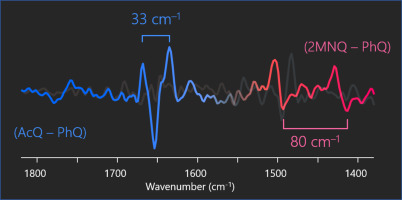
Infrared absorption bands associated with the neutral state of quinones in the A1 binding site in photosystem I (PSI) have been difficult to identify in the past. This problem is addressed here, where time-resolved step-scan FTIR difference spectroscopy at 77 K has been used to study PSI with six different quinones incorporated into the A1 binding site. (P700+A1- - P700A1) and (A1- - A1) FTIR difference spectra (DS) were obtained for PSI with the different quinones incorporated, and several double-difference spectra (DDS) were constructed from the DS. From analysis of the DS and DDS, in combination with density functional theory based vibrational frequency calculations of the quinones, the neutral state bands of the incorporated quinones are identified and assigned. For neutral PhQ in the A1 binding site, infrared absorption bands were identified near 1665 and 1635 cm-1, that are due to the C1O and C4O stretching vibrations of the incorporated PhQ, respectively. These assignments indicate a 30 cm-1 separation between the C1O and C4O modes, considerably less than the ~80 cm-1 found for similar modes of PhQ-. The C4O mode downshifts due to hydrogen bonding, so the suggestion is that hydrogen bonding is weaker for the neutral state compared to the anion state, indicating radical-induced proton dynamics associated with the quinone in the A1 binding site in PSI.
中文翻译:

用于研究光系统A1结合位点中醌的时间分辨FTIR差异光谱法I:鉴定中性态醌带。
过去,很难识别与光系统I(PSI)中A1结合位点中的醌中性状态相关的红外吸收带。此问题已在此处解决,其中使用时间分辨的阶跃扫描FTIR差异光谱在77 K的条件下研究PSI,其中将六个不同的醌掺入A1结合位点。获得了掺入不同醌的PSI的(P700 + A1--P700A1)和(A1--A1)FTIR差异光谱(DS),并从DS构造了几个双差光谱(DDS)。通过对DS和DDS的分析,结合基于醌的基于密度泛函理论的振动频率计算,可以识别并指定所掺入醌的中性能带。对于A1结合位点的中性PhQ,分别在1665和1635 cm-1附近发现了红外吸收带,这分别是由于所结合的PhQ的C1O和C4O拉伸振动所致。这些分配表明C1O和C4O模式之间的间隔为30 cm-1,大大小于类似PhQ-模式的〜80 cm-1。由于氢键,C4O模式下移,因此建议中性状态的氢键比阴离子状态弱,这表明自由基诱导的质子动力学与PSI中A1结合位点的醌有关。
更新日期:2020-03-22
中文翻译:
用于研究光系统A1结合位点中醌的时间分辨FTIR差异光谱法I:鉴定中性态醌带。
过去,很难识别与光系统I(PSI)中A1结合位点中的醌中性状态相关的红外吸收带。此问题已在此处解决,其中使用时间分辨的阶跃扫描FTIR差异光谱在77 K的条件下研究PSI,其中将六个不同的醌掺入A1结合位点。获得了掺入不同醌的PSI的(P700 + A1--P700A1)和(A1--A1)FTIR差异光谱(DS),并从DS构造了几个双差光谱(DDS)。通过对DS和DDS的分析,结合基于醌的基于密度泛函理论的振动频率计算,可以识别并指定所掺入醌的中性能带。对于A1结合位点的中性PhQ,分别在1665和1635 cm-1附近发现了红外吸收带,这分别是由于所结合的PhQ的C1O和C4O拉伸振动所致。这些分配表明C1O和C4O模式之间的间隔为30 cm-1,大大小于类似PhQ-模式的〜80 cm-1。由于氢键,C4O模式下移,因此建议中性状态的氢键比阴离子状态弱,这表明自由基诱导的质子动力学与PSI中A1结合位点的醌有关。


 京公网安备 11010802027423号