Our official English website, www.x-mol.net, welcomes your
feedback! (Note: you will need to create a separate account there.)
In vivo labeling of epithelial cell-associated antigen passages in the murine intestine.
Lab Animal ( IF 5.9 ) Pub Date : 2020-02-10 , DOI: 10.1038/s41684-019-0438-z Kathryn A Knoop 1, 2 , Devesha H Kulkarni 1 , Keely G McDonald 1 , Jenny K Gustafsson 3 , Jazmyne E Davis 1 , Alexandria N Floyd 1 , Rodney D Newberry 1
Lab Animal ( IF 5.9 ) Pub Date : 2020-02-10 , DOI: 10.1038/s41684-019-0438-z Kathryn A Knoop 1, 2 , Devesha H Kulkarni 1 , Keely G McDonald 1 , Jenny K Gustafsson 3 , Jazmyne E Davis 1 , Alexandria N Floyd 1 , Rodney D Newberry 1
Affiliation
|
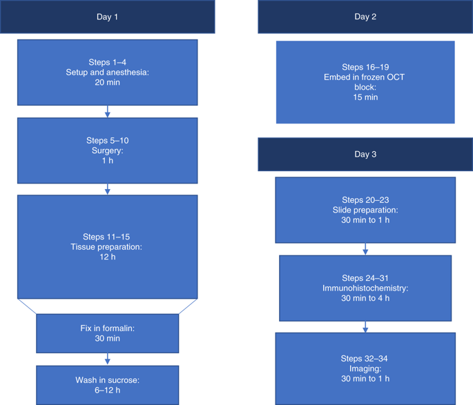
The intestinal immune system samples luminal contents to induce adaptive immune responses that include tolerance in the steady state and protective immunity during infection. How luminal substances are delivered to the immune system has not been fully investigated. Goblet cells have an important role in this process by delivering luminal substances to the immune system through the formation of goblet cell-associated antigen passages (GAPs). Soluble antigens in the intestinal lumen are transported across the epithelium transcellularly through GAPs and delivered to dendritic cells for presentation to T cells and induction of immune responses. GAPs can be identified and quantified by using the ability of GAP-forming goblet cells to take up fluorescently labeled dextran. Here, we describe a method to visualize GAPs and other cells that have the capacity to take up luminal substances by intraluminal injection of fluorescent dextran in mice under anesthesia, tissue sectioning for slide preparation and imaging with fluorescence microscopy. In contrast to in vivo two-photon imaging previously used to identify GAPs, this technique is not limited by anatomical constraints and can be used to visualize GAP formation throughout the length of the intestine. In addition, this method can be combined with common immunohistochemistry protocols to visualize other cell types. This approach can be used to compare GAP formation following different treatments or changes to the luminal environment and to uncover how sampling of luminal substances is altered in pathophysiological conditions. This protocol requires 8 working hours over 2-3 d to be completed.
中文翻译:

小鼠肠道中上皮细胞相关抗原通道的体内标记。
肠道免疫系统对管腔内容物进行采样以诱导适应性免疫反应,包括稳态耐受和感染期间的保护性免疫。尚未充分研究管腔物质如何传递到免疫系统。杯状细胞通过杯状细胞相关抗原通道 (GAP) 的形成将管腔物质输送到免疫系统,在这一过程中发挥着重要作用。肠腔中的可溶性抗原通过 GAP 跨细胞转运穿过上皮细胞并递送至树突状细胞以呈递给 T 细胞并诱导免疫反应。通过使用形成 GAP 的杯状细胞吸收荧光标记的葡聚糖的能力,可以识别和量化 GAP。这里,我们描述了一种可视化 GAP 和其他细胞的方法,这些细胞通过在麻醉下的小鼠中腔内注射荧光葡聚糖、组织切片用于幻灯片制备和荧光显微镜成像,从而能够吸收腔内物质。与以前用于识别 GAP 的体内双光子成像相比,该技术不受解剖学约束的限制,可用于可视化整个肠道长度的 GAP 形成。此外,这种方法可以与常见的免疫组化协议相结合,以可视化其他细胞类型。这种方法可用于比较不同治疗或改变管腔环境后的 GAP 形成,并揭示在病理生理条件下如何改变管腔物质的采样。
更新日期:2020-02-10
中文翻译:
小鼠肠道中上皮细胞相关抗原通道的体内标记。
肠道免疫系统对管腔内容物进行采样以诱导适应性免疫反应,包括稳态耐受和感染期间的保护性免疫。尚未充分研究管腔物质如何传递到免疫系统。杯状细胞通过杯状细胞相关抗原通道 (GAP) 的形成将管腔物质输送到免疫系统,在这一过程中发挥着重要作用。肠腔中的可溶性抗原通过 GAP 跨细胞转运穿过上皮细胞并递送至树突状细胞以呈递给 T 细胞并诱导免疫反应。通过使用形成 GAP 的杯状细胞吸收荧光标记的葡聚糖的能力,可以识别和量化 GAP。这里,我们描述了一种可视化 GAP 和其他细胞的方法,这些细胞通过在麻醉下的小鼠中腔内注射荧光葡聚糖、组织切片用于幻灯片制备和荧光显微镜成像,从而能够吸收腔内物质。与以前用于识别 GAP 的体内双光子成像相比,该技术不受解剖学约束的限制,可用于可视化整个肠道长度的 GAP 形成。此外,这种方法可以与常见的免疫组化协议相结合,以可视化其他细胞类型。这种方法可用于比较不同治疗或改变管腔环境后的 GAP 形成,并揭示在病理生理条件下如何改变管腔物质的采样。


 京公网安备 11010802027423号